Diabetic Retinopathy (DR)
Retinal disease classified by microvascular and neuronal complications of diabetes.
Wan, T., Li, X., Sun, Y., Li, Y., & Su, Y. (2015). Recent advances in understanding the biochemical and molecular mechanism of diabetic retinopathy. Biomedicine & Pharmacotherapy, 74, 145-147. doi:10.1016/j.biopha.2015.08.002
Epidemiology,
At a Glance...
Bhavsar, A. (2015, April 1). Diabetic Retinopathy. Retrieved November 25, 2015, from http://emedicine.medscape.com/article/1225122-overview#showall
Paulus, Y., & Ray, G. (2009). Diabetic Retinopathy: A growing concern in an aging population. Geriatrics, 64(2), 16-26.
Leading cause of irreversible blindness in working-age Americans.
Strong correlation between duration of diabetes and development of DR.
DR development is rare within the first 5 years of onset of diabetes, and risk increases thereafter.
Effects approximately 700,000 persons in the US, with an annual incidence of 65,000 cases.
Prevalence of 28.5% in diabetic patients aged 40+
Responsible for approximately 8,000 eyes becoming blinder each year, and is therefore, responsible for 12% of all blindness.
Increased risk for patients of Native American, Hispanic, and African American heritage
Disease Time Course
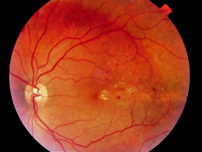
Early Stages: Non-Proliferative Diabetic Retinopathy (NPDR)
Hypoglycemia-induced weakening of retinal capillaries resulting in leakage of blood and fluid into retinal pigment epithelium.
(Wan, 2015)
Symptoms
Asymptomatic
Diagnoses
Microaneurysms
Earliest clinical manifestation of DR resulting from diabetes-induced vascular changes such as endothelial proliferation and pericyte damage, which leads to capillary out-pouching and subsequent aneurysm formation.
Dot & Blot Hemorrhages
Microaneurysms rupture at various layers causing blood leakage into retina.
Retinal Edema & Hard Exudates
Ruptured capillaries allow fluids and protein to accumulate in retina, causing it to sweep. Hard exudates (yellow spots) are lipid residue that are left behind on retina.
Treatment
Laser Photocoagulation
Laser is used to destroy/shrink abnormally, weakened blood vessels in retina.
Advanced Stages: Proliferative Diabetic Retinopathy (PDR)
Retinal ischemia stimulates proliferation of new, equally weak blood vessels which lead to severe conditions and vision loss.
Diabetic Retinopathy - Topic Overview. (2014, June 1). Retrieved November 25, 2015, from http://www.webmd.com/diabetes/tc/diabetic-retinopathy-topic-overview
Symptoms
Blurred, double, distorted vision
Floaters
Partial or total loss of vision
Diagnoses
(Bhavsar, 2015)
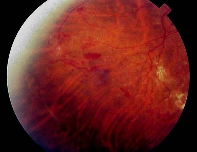
Neovascularization
Hallmark of PDR. Retinal ischemia stimulates growth of new, weak retinal capillaries, which subsequently develop microaneurysms and hemorrhage.
Retinal Detachment
Resulting from scar tissue pulling on the retina
Vitreous Hemorrhaging
Blood clots escaping into vitreous humor
Macular Edema
Retinal edema that occurs centrally effects the macula, resulting in macular edema - the leading cause of vision loss in diabetics
Neuronal Complications
(Wan, 2015)
Reduced corneal nerve sensations
Impaired autonomic innervation to pupil
Altered functionality of retinal sensory nerve
Treatment
(WebMD, 2014)
Vitrectomy
Surgical removal of vitreous gel in cases of severe scar tissue formation, vitreous hemorrhaging, or retinal detachment.
VEGF
Medications that shrink the proliferating capillaries. Used in cases of long-standing vitreous hemorrhaging or retinal detachment.
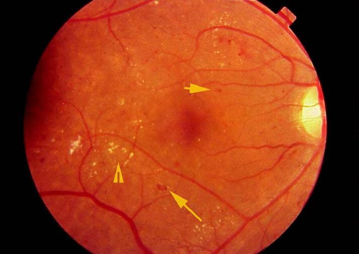
Retinal findings in background diabetic retinopathy, including blot hemorrhages (long arrow), microaneurysms (short arrow), and hard exudates (arrowhead).
(Bhavsar, 2015)