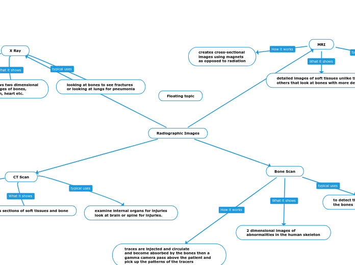
Radiographic Images
X Ray
a machine sends individual X-ray
particles called photons through
the body producing photos
shows two dimensional
images of bones,
teeth, heart etc.
looking at bones to see fractures
or looking at lungs for pneumonia
CT Scan
the machine takes a series of x-ray views from many angles and are then combined to make cross sectional images
cross sections of soft tissues and bone
examine internal organs for injuries
look at brain or spine for injuries.
MRI
creates cross-sectional
images using magnets
as opposed to radiation
detailed images of soft tissues unlike the others that look at bones with more detail
monitor treatment for conditions in the chest abdomen and pelvis and look at organs more closer.
Bone Scan
traces are injected and circulate
and become absorbed by the bones then a gamma camera pass above the patient and pick up the patterns of the tracers
2 dimensional images of
abnormalities in the human skeleton
to detect the spread of metastatic cancer in the bones