Membrane Bound Organelles
Nuclear envelope- double membrane enclosing the nucleolus
Cytoplasm- surrounds the organelles
Plasma Membrane-
membrane enclosing the cell; serves as a barrier between the cell, things outside of the cell, and things that want to enter the cell
Consists of double layer of phospholipids with various proteins attached to or embedded in it
Membrane Transport and Structure
Phospholipid Bilayer
Amphipathic
Membrane Proteins
Different Functions of Membrane Proteins
Transmembrane (Integral) Proteins
- proteins that are located within the plasma
membrane
- composed of alpha helices, N-terminus outside and C-Terminus inside cell
Peripheral Proteins
- proteins located on the outside
of the plasma membrane
Transport Proteins
and Membrane Transport
Selective Permeability
- regulates cell movement, cell traffic, and inter/extracellular interactions
Passive Transport
- Type of transport that doesn't require ATP
- High to Low
- Down Concentration Gradient
Active Transport
- Type of transport that REQUIRES ATP
- Low to High
- Against Concentration Gradient
Pumps
Electrogenic Groups
CoTransport
Bulk Transport
Enzymatic Activity
- Carries out metabolic processes
- Membranes have active sites and chemical receptors that carry out sequential steps
Signal Transduction
- Possess certain receptors that are fit to receive messages from signaling molecules
- Molecules binds to cytoplasmic proteins which have ends that are outside and inside the cell.
Cell-Cell Recognition
- Glycoproteins serve as ID tags which are recognized by certain proteins and receptors of membrane cells
- Short-Lived Connection
Intercellular Joining
and Cell Junctions
- Membranes cells hook together in junctions
- Long-Lived Connection
Attachment to ECM and Cytoskeleton
Connects to the cytoskeleton's
microfilaments non-covalently
The connection to the ECM helps
cell perform inter/extracellular changes
Endoplasmic reticulum-network of membranous sacs and tubes; active in membrane synthesis and other
Rough ER-
ribosomes are attached
Smooth ER
Golgi apparatus- organelle active in synthesis. modification, sorting, and secretion of cell products
cis face, same side, is located near ER
trans face, opposite side is located gives rise to vesicles that pinch off and travel to other sides^
Lysosomes-digestive organelle where macromolecules are hydrolyzed
carry out intracellular digestion in a variety of circumstances
Cytoskeleton- reenforces cell shape; functions in cell movement; components are made of proteins
Structures and functions of Cytoskeleton
Microtubules
Microfilaments
Intermediate filaments
Mitochondrion- cellular respiration occurs and ATP is generated
Produces ATP which is energy that the cell uses
outer membrane is smooth, but the inner membrane is convoluted, with folding called cristae; there are two compartments
intermembrane space- between the inner and outer membranes
mitochondrial matrix-
compartment of the mitochondrion containing enzymes and substrates for the citric acid cycle, as well as ribosomes and DNA
Cellular Respiration
1. Glycolysis
(Steps 1 and 3)
- occurs in the cytoplasm
- Energy Investment and Energy Pay Off Phases
- ATP formed with Substrate-Level Phosphorylation (sLP)
INPUTS
- 1 Glucose
- 2 NAD+
- 2 ATP
OUTPUTS
- 2 Pyruvate
- 2 NADH
- 4 ATP
2. Pyruvate Oxidation
- occurs from the cytosol to the Mitochondrial Matrix
INPUTS
- 2 Pyruvate
- 2 CoA
- 2NAD+
OUTPUTS
- 2 Acetyl CoA
- 2 NADH
3. Citric Acid Cycle
(steps 1 and 3)
- occurs in the Mitochondrial Matrix
- ATP formed with SLP
INPUTS
- 2 Acetyl CoA
- 6 NAD+
- 2 FAD
OUTPUTS
- 2 ATP
- 6 NADH
- 2 FADH2
4. Oxidative Phosphorylation
- occurs among the Mitochondrial Matrix, Inner Mitochondrial Membrane, and the Inner-membrane Space
Electron Transport Chain
- NADH: I, Q, III, Cyt C, and IV
- FADH2: II, Q, III, Cyt C, and IV
- Oxygen is the final electron acceptor
As electrons move across the complexes, H+ is pumped out into the intermembrane space establishing a proton gradient.
Chemiosmosis
The proton gradient coming back into the organelle through ATP Synthase produces ATP. Gradient spins motor that attaches the inorganic phosphate to ADP to create ATP.
The ETC and Chemiosmosis are coupled to generate ATP. The gradient from ETC is required to produce ATP with ATP Synthase.
INPUTS
- O2
- 10 NADH
- 2 FADH2
OUTPUTS
- H2O
- 26-28 ATP
Microvilli- increase the cells surface area
Peroxisome- organelle with various specialized metabolic functions; produces hydrogen peroxide as a by-product and then converts it to water
Flagellum- motility structure present in some animal cells, composed of a cluster of microtubules within an extension of the plasma membrane
Centrosomes- region where the cell's microtubules are initiated; contains a pair of centrioles
Ribosomes
Endomembrane system- membranes of this system are related either through direct physical continuity of transfer by vesicles
Extracellular Matrix (ECM)
Specific to animal cells as plants have cell walls
Carbohydrates
Lipids
Proteins
Nucleic Acids
DNA (deoxyribonucleic acid)
provides own information for replication
directs synthesis for mRNA and controls protein synthesis
gene expression
translation (info from mRNA makes proteins)
transcription (info from DNA is used to make RNA)
RNA (ribonucleic acid)
adenine, cytosine, uracil, and guanine
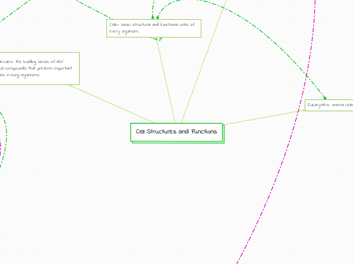
Organelles
Fimbriae- Bacteria can attach to particular receptor structures thanks to attachment structures.
Nucleiod- DNA situated here that is not membrane-enclosed
Cytoplasm- contains organic molecules and enzymes and is primarily composed of water.
Cell wall- rigid, external to the plasma membrane, provides security and shape
Glycocalyx- An outer layer is made up of a slime or capsule layer.
conserves fluids and guards the cell from being swallowed by other organisms. fights off phaagocytosis
Plasma Membrane- Selectively The cytoplasm is enclosed and controls the flow of substances during nutrition and waste transport as well as the location of metabolic processes.
Ribosomes-protein production and synthesization
Producers
Autotroph
Photoautotroph
Organism types:: Photo synthetic prokaryotes algae
Energy comes from a light carbon source CO2 HCO3
Chemoautotroph
Energy comes from in organic chemicals carbon source CO3 HCO3
Heterotroph
Photoautotroph
the sources are Light and organic compounds
Chemoautotroph
Organic Compounds
a nucleoside DOES NOT have a phosphate group
purines (A and G)
2 carbon nitrogen ring bases
pyrimidines (C, T, and U in RNA)
1 carbon nitrogen ring bases
joined by condensation/dehydration
reactions= phosphodiester bond/linkage
Functions
Caries out the transduction part of the signaling pathway
Most other intracellular receptors function in the same way
Details
1. Found in the cytoplasm or nucleus of target cell
2. Signaling molecule passes through target cell's plasma membrane
Steroid hormone interactions
1.The steroid hormone passes through the plasma membrane
2. Hormone binds to a receptor protein in the cytoplasm activating it
3. The hormone-receptor complex enters the nucleus and binds to specific genes
1.The hormone receptor complex turns on genes in a cells DNA function by being transcribed and processed into messenger RNA in the nucleus
2. Leaves the nucleus and is translated into specific proteins by ribosomes in the cytoplasm
3. Proteins called transcription factors control with genes are tuned on/ transcribed in mRNA
4. The bound proteins acts as a transcription factor , stimulating the transcription of the gene into mRNA
5. The mRNA is translated into a specific protein
G Protein linked Receptor
1. First messenger binds to GPCR, activating it
2. Activated GPCR binds to G protein, which is then bound by GTP, activating G protein
3. Activated G protein/GTP binds to adenylyl cyclase. GTP is hydrolyzed, activating adenylyl cyclase
4. Activated adenylyl cyclase converts ATP to cAMP
5. cAMP (second messenger) activates another protein, leading to cellular responses
Tyrosine Kinase Receptor
(made of 2 polypeptides
which dimerize when a
signal molecule is bound to
each polypeptide)
1. Signal molecule binds the each of the binding-sites
2. Polypeptides dimerize (join together), activating them (unphosphorylated dimer)
3. Autophosphorylation (phosphate group taken from ATP and added to the other polypeptide)
4. Fully activated receptor tyrosine-kinase (phosphorylated dimer)
5. Activated receptor now interacts with other relay proteins to bring out cellular response 1/2
Ion Channel Receptor
(acts as a gate for ions
when the receptor changes
shape)
gate allows specific ions (Na+ or Ca+) through a channel
very important in the nervous system
movement of ions in these channels may change voltage across membranes = triggers action potential
Action potential-
electrical signal that propagates along the membrane of a neuron or other excitable cells as a nongraded depolarization
COMPONENTS
m and tRNAs | Ribosomal Components:
E, P, A sites | large and small subunits | peptidyl transferase, amino-acyl tRNA synthetase | stop, start codons and anticodons | initiation, elongation, and release factors
PROCESS
:1. INITIATION:
- mRNA, tRNA, two ribosomal subunits come together
- GTP and initiation factors required to assemble this complex
- Small ribosome and tRNA bind to 5’cap, scans mRNA for AUG, and then large subunit assembles to form translation initiation complex
2. ELONGATION
- tRNA to A site with amino acid, moves to P site to for peptide bonds, then tRNA exits at E site
- peptide chain grows at the P site
- Each step GTP is used with GDP+P remaining
- Amino acids are added from N(amino) to C (carboxyl)
- peptidyl transferase creates the peptide bonds
- amino-acyl tRNA synthetase adds the appropriate amino acid after reading the mRNA codon
3. TERMINATION
- Stop codon read, release factors sits at A site, and then breaks apart the complex
- Driven by GTP
POST-TERMINATION
- Endomembrane System If it has an ER signal sequence, free ribosomes to the ER to the golgi body, out the cell/other locations via vesicles
- Amino acid signal sequence tell proteins where to go
- Secretory Pathway: path take protein from synthesis to modification to secretion
- Glycoproteins formed from glycosylation, (process of adding carbohydrate groups to proteins)
- Protein destinations: mitochondria, chloroplast, peroxisomes, nucleus
PROKARYOTES VS. EUKARYOTES
PROKARYOTES
- Occurs in the cytoplasm and uses mRNA and tRNA
- Ribosomal components: 70S, Large subunit: 50S (31 proteins) and Small subunit: 30S (21 proteins)
- All steps of translation the same, but elongation uses formal-MET, and lack of organelles means protein destinations are cytosol or plasma membrane
EUKARYOTES
- starts at free ribosomes (cytosol) but moves to
ER if initiated
- Ribosomal components: 80S, Large subunit: 60S (~ 45 proteins) and Small subunit: 40S (~ 33 proteins)
- these have more possibilities with more organelles and pathways than prokaryotes, and it just uses MET
Prokaryotes vs. Eukaryotes
Prokaryotes
-Occurs in cytoplasm, allows coupling between translation and transcription, means as the mRNA made can immediately be translated
-Only RNA polymerase
Eukaryotes
-occurs in nucleus, pre-mRNA is formed
-different RNA polymerases are used to make different RNAs, focus on RNA polymerase II
- initiation uses transcription factors that bind to promoter before RNA polymerase II can bind and uses the transcription initiation complex
-At termination 5' end a CAP is added, and at 3' end a polyA tail is added with the help of polyA polymerase
Procces
Initiation
1. RNA polymerase bind to DNA on the upstream at start site called promoter and then uses a DNA strand as a template strand
2. DNA strand unwinds, polymerase begins to transcribe at start site
3. RNA polymerase make new strand from 5' to 3' but read it from 3' to 5'
Elongation
1. Polymerase moves down stream, unwinding DNA as it transcribes
2. Polymerase add nucleotides to the 3' end of growing RNA molecule
3. New RNA molecule peels away from DNA template and the DNA double helix re-forms
Termination Eukaryotes
1. RNA polymerase II transcribe a signal sequence of AAUAAA to cut RNA transcript free from polymerase
2. pre-mRNA is released then undergoes processing
Termination Prokaryotes
1. The polymerase detaches from DNA and releases then transcript with the help of a transcribed terminator
Transcription Factors
-Eukaryotes use these in initiation the transcription factor bind to promoter before polymerase II
- forms transcription initiation complex
Eukaryotes RNA modifications
RNA Processing
1. ends or pre-mRNA are modified; the 5' end receives a cap and the '3 end receives a poly-A tail
- helps with export of mature mRNA from nucleus, help protect from degradation and help ribosomes attach to 5' end in cytoplasm
RNA Splicing
1. Introns are cut out of molecule and exons are joined together with the help of a complex called spliceosome
Structure made up of...
Nucleotides (connected together through condensation/dehydration reactions & held together by hydrogen bonds)
Composed of...
Phosphate Group
Sugar (deoxyribose)
Nitrogenous Base
Start of Replication...
separate 2 strands of DNA at ORI (origin of replication) sequences and form a bubble
- Helicase separates strands
-SSB (single stranded proteins) keep DNA apart
-Topoisomerase relieves strain on DNA
-Primase synthesizes RNA Primers
- DNA polymerase 3 adds nucleotides to 3'
(needs the help of another protein-sliding clamp-keeps DNA on parent strand)
Synthesis of leading strand
-DNA POlymerase 3 synthesizes
moving toward replication fork (5' to 3')
Synthesis of lagging strand
-multiple RNA primers are laid out, DNA pol 3 extend, okazaki fragments are formed
-Dna Polymerase 1 removes RNA and replaces it with DNA nucleotides
-ligase seals gaps
cytosol^
mitochondriion
Perixsome
chloroplast
nucleus
Endoplasmic Reticulam
Golgi Apparatus
cell extersior
plasma membrane
lusosomes
other parts of end membrane system
In the cytosol polypeptide synthesis starts on a free ribosome
A signal peptide is bound by a signal recognition particle (SRP), the synthesis is momentarily stopped
A receptor in the ER membrane is where SRP binds
Polypeptide synthesis resumes when SRP leaves
A receptor protein complex enzyme splits the signal peptide
After leaving the ribosome, the finished polypeptide assumes its final configuration
Becomes membrane protien
selected outside the cell
Glycosylation
Glycoprotein (Carbohydrates and Proteins)
Secondary Proteins
Amylase is released by digestive enzymes
Insulin is secreted by peptide hormones
Casein is secreted by milk proteins
Albumin is secreted by serum protiens
Proteins from extracellular matrix secrete college
additional locations