Organ System Concept Map
Wrap Up!
Respiratory System
Structural Plan
Leaves+ Alveoli with the microscopic sacs enclosed by networks of capillaries
Lower Respiratory tract
Trachea, Bronchial tree, and lungs
Trachea (windpipe)
Structure
Tube about 11cm (4.5) inches long
Extends from the larynx into the thoracic cavity
C-shape rings of cartilage hold trachea open
Function
Passageway for air to move to and from lungs
Obstruction
Blockage of trachea occludes the airway and if blockage complete causes death in minutes
Tracheostomy- emergency surgery done to bring airflow back into the respiratory system.
Bronchi, Brolchioles, and Alveoli
Structure
Trachea branaches into right and left bronchi
Each Bronchus branches into bronchioles
Bronchioles end in clusters of microscopic albeolar sacs
Wakks of these sacs are made up of alveoli
Function:
Bronchi and bronchioles- air distribution
Alveoloi- exhcnage of gases between air and blood
Exchanges of gas in lungs
CO2 from blood into alveoli and out of the body
O2 from air into the alveoli then into blood
Exchange of gases in tissues
02 from blood into the tissue
CO2 from tissue into blood
The cellular level the gas is going straight to the blood that’s passing by from the alveoli straight to the blood that’s passing by from the alveoli across to the capillary with blood inside and so they’re just showering in the direction of gas floor from the red blood cells and so on.
Think of the respiratory system asa n inverted hollowed out tree
Upper Respiratory Tract
Nose, pharynx, and larynx
Nose
Structure
Nasal Septum
Mucous membrane lines the nose
Sinuses drain into nose
Functions
Wamrs and mositents inhaled air
Contains sense organs of smell
Larynx
Structure
Framework of several pieces of cartilage
Thyroid Cartilage
Lungs
Structure:
Fills chest cavity (with heart and blood vessels
Apex- narrow upper part of each lung under collarbone
Base- Broad lower part of each lung rests on diaphragm
Pleura- moist smooth slippery membranes that line chest cavity and cover outer surface of lungs
Reduces friction between the lungs and chest wall during breathing
Friction
Breathing (Pulmonary ventilation
Types of Breathing
Eupnea- normal breathing
Hyperventilation- rapid and deep respirations/ caused by physical and emotional stimuli (Stroke, Anxiety, etc.).
More CO22 will be removed from the blood stream than the body can produce
Hypoventilation- slow and shallow respirations
Inadequate gas exchange causes increased concentrations of CO2
Many causes stroke holding breath opiate drugs etc.
Dyspnea- labored or difficult respirations
Apnea- stopped respirations
Respiratory Arrest- Failure or resume breathing after a period of apnea.
Respiratory Mucosa
Specialized membrane that lines the air distribution tubes in the respiratory tree
125 m; of mucous produced each day
Forms a mucus blanket
Traps inspired irritants
Cilia beat in only one direction
Moves mucus upward to pharaynx for removal
Pharaynx (Throat)
Structure
About 12.5com 5 inches long
Two nasal cavities, mouth, esophaguse, larynx, and autidotry tubes all have openings into the pharaynx
Mucus membrane lines the pharaynx
Functions:
Passageway for food and liquids
Air distrubtion passageway for air
Epiglottis
Mucous lining
Vocal cords stretch across interior of the larynx
Functions
Air distribution, Voices Production
Respiration
Mechanics of breathing
Two phases
Inhalation
Expiration
Changes in size and shape of thorax cause changes in air pressure within that cavity in the lungs
Air pressure differences actually cause air to move into and out of the lungs
Inspiration-
Active process- air moves into the lungs
Muscles are required
Diaphragm flattens during inspiration—increases top-to-bottom length of thorax
External intercostals contraction expands the ribs
Increases in the size of the chest cavity reduces pressure within it air then enters the lungs
Expiration
A passive process
Thorax returns to its resting size/shape
In forceful expiration, muscles used are:
Internal intercostals-contraction depresses the rib cage
Abdominal muscles elevate the diaphragm
Reduction in the size of the thoracic cavity increases its pressure and air leaves the lungs
Simialrites and Correlations
Both Organ systems produce movement throughout the body.
During inhalation the muscles in the respiratory system open up and extend along with tension on the surface to allow maximum surface area to bring in the most air.
During Exhalation the lungs sink like when a balloon runs out of air.
The respiratory system and the muscular system are both voluntary and involuntary as you can hold your breath and exhale your breath and your body keeps your body breathing. Along with the muscles that work voluntarily and involuntarily. The muscle movements allow there to be breathibg mechanisms Involving with the diaphgram and the intercostals
The Respiratory System and The Muscular system work hand in hand in helping carbon mateirals such as carbon dixodie and other fluids move throughout the body like the digestive system moves food around using peristalsis the body uses it to move the air and msucular systems throughout the body
The respiratory systm is needed for the muscular system to function because the muscles of the skeletal system need the lungs and the respriatory system to give the tissue nutrients to move.
The esophagus shares skeletal muscle tissue to protect the lungs and the trachea from getting injured due to pollutants and irritants.
For muscles to function they need to recieve oxygen from the respiratory system. As working muscles produce gaseous wastes which are carried by the blood back to the respiratory system were they are carried out of the body
https://socratic.org/questions/how-does-the-respiratory-system-depend-on-the-muscular-system#:~:text=Explanation%3A,the%20respiratory%20system%20and%20expelled.
https://my.clevelandclinic.org/health/articles/21205-respiratory-system
https://www.visiblebody.com/blog/anatomy-and-physiology-the-relationships-of-the-respiratory-system
Musclar System
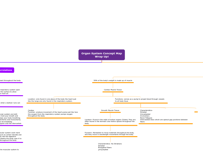
50% of the body's weight is made up of muscle
Cardiac Muscle Tissue
Location: only found in one place of the body the heart just like the lungs are only found in the respiratory system.
Cardiac
Function: produce movement of the heart pump just like how the oxygen from the respiratory system pumps oxygen throughout the body.
Functions- serves as a pump to propel blood through vessels to all body tisue
Smooth Muscle Tissue
Location: Found in the walls of hollow organs (Tubes) They are often found in the stomach and hollow spaces throughout the body.
Function: Peristalisis to move materials throughout the body, and they move in wavelength movements through the body.
Characteristics- No Striations
Striated
Elongated Fibers
unnucleated
Involuntary
Smooth muscle function: it’s to move materials through the body.
Characteristics-
Striated
Unnueleated
Involuntary
Never Fatigues
Intercalated discs whcih are speical gap junctions between fibers
The muscles in our body are made up of water and proteins. Just like the nutrients that are in the bloodstream.
Skeletal Muscle Tissue
Muscle Tissue- is the only body tissue able to contract or shorten. The largest Muscle in the body is the gleutus maximus. Muscular tissue goes through the whole body like how there are veins running through the body.
Skeletal Muscle organization
You have individual muscle cells and fibers called the Perimysium. This is like the individual hair roots that are on the head
The bunches of muscle cells and fibers that are wrapped in branches are called the Fasicles. This includes many bundles of wire put together called fasicles
The bunches of individual muscle fibers are wrapped in Perinysium. The fasicles are inside this strong protective connective tissue. That is made of steel like strong protective covering.
The Final Protective Layer that goes around the whole muscular Fiber is the epimysium it wraps around the whole connective tissue fibers that are bunched together.
Skeletal also:
Maintains posture
Stabilizes joints
Generates heat – when you contract your skeletal muscles when you d been outside I. The cold and jog in place it warms you up and when you exercise you get warmer and you settle and it is the contraction of all of the muscles and regulates body temp
A byproduct of as a result of contraction
Mainstrains body temp
Ex: shivering, exercising
The Fasicles are wrapped in a thin connective tissue called an endoymysium along with each indivudal are wrapped in endyomysium
Each muscle fiber could be a piece of spaghetti uncooked spaghetti and then when you put all those prices of spaghetti together into a fascial the box or plastic you know thing that you put all those sticks of spaghetti in could be the perimysium. Which stops each fascicle and then if you have a box that contains many boxes of spaghetti that the outer box could be the epimysium
skeletal muscles Function: to move the skeletal system and bones and joints.
The epimysium is thicker, and it covers then entire muscle. A bunch of fascicles and wraps it around and then it becomes continuous it’s a continuous membrane or tissue that becomes a tendon which is a really tough connective tissue so it’s continuous with this structure, so it becomes a tendon which connects the muscle to the bone and um the other
Location: Cover the skeletal framework just like the skin covers the skeletal.
Function: Movment of the Skeleton, which is when the body is in motion.
The tissues are striated and are long fibers that create tendons and are voluntary movemnts such as when you are walking and when you are exercising it can cause tiredness with the muscles.
Characteristics-
striated
elongated fibers
multinucleated
voluntary
can fatigue
Subtopic