Cytoskeleton: reinforces cell shape; functions in cell movement; components are made of proteins
Microfilaments
Intermediate filaments
Microtubules
Lysosome: digestive organelle where macromolecules are hydrolyzed
Nucleus: carries genes, regulates functions, holds structures that contain hereditary information, contains chromosomes
Nuclear Envelope: double membrane enclosing the nucleus; perforated by pores; continuous with ER
Nucleolus: non membranous structure involved in production of ribosomes; a nucleus has one or more nucleoli
Chromatin: material consisting of DNA and proteins visible in a dividing cell as individual condensed chromosomes
Golgi Apparatus: organelle active in synthesis, modification, sorting, and secretion of cell products
Mitochondria: where cellular respiration occurs and most ATP is generated
Ribosomes: make proteins; can be free in cytosol or bound in rough ER/nuclear envelope
Ribosomes: protein synthesis
Flagellum: motility structure present in some animal cells; composed of cluster of microtubules within an extension of plasma membrane
Centrosome: region where the cell's microtubules are initiated; contains a pair of centrioles
Microvilli: projections that increase the cell's surface area
Peroxisome: organelle with various specialized metabolic functions; produces hydrogen peroxide as a by-product then converts it to water.
Endoplasmic Reticulum: network of membranous sacs and tubes; active in membrane synthesis and other metabolic processes.
Smooth ER
Rough ER (ribosome studded)
Chloroplast: photosynthetic organelle; converts energy of sunlight to chemical energy stored in sugar molecules
Plasmodesmata: cytoplasmic channels through cell walls that connect the cytoplasm of adjacent cells.
Central Vacuole: prominent organelle in older plant cells; functions include storage, breakdown of waste products, and hydrolysis of macromolecules, enlargement of vacuole is essential to cell growth
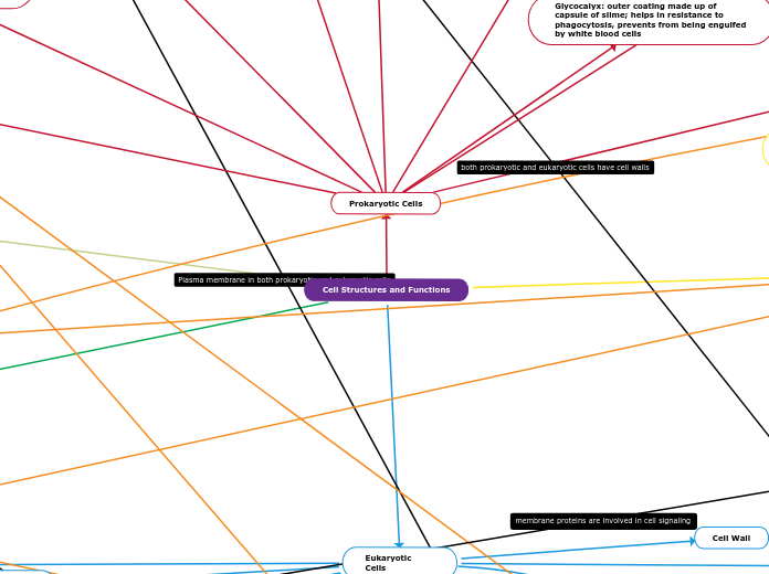
Cell Wall
Quaternary Structure
When two or more polypeptides come together to form a functional protein. Mostly weak interactions such as hydrogen bonding hold the subunits together to form quaternary structures.
Tertiary Structure
Formed when the polypeptide folds into a 3D shape through the interaction of R groups. R-group interactions involve hydrogen and ionic bonding, dipole-dipole and hydrophobic interactions. Also where disulfide bonds occur.
Secondary Structure
The main chain parts interact through hydrogen bond formations. This results in alpha helices or beta pleated sheets.
Primary Structure
A sequence of amino acids held together by peptide bonds. One amino end and one carboxyl end.
Hydrophilic Head
Since the heads are hydrophilic, they face outward and are attracted to the intracellular and extracellular fluid.
Hydrophobic Tail
the type of hydrocarbon tail in phospholipids also affects the fluidity of the plasma membrane
unsaturated hydrocarbon tails with kinks are not as tightly packed so they are more fluid than the tightly packed saturated hydrocarbon tails that do not have kins
Since the tails are hydrophobic, they face the inside, away from the water and meet in the inner region of the membrane.
Those regions of the protein that must interact with the strongly hydrophobic center of the lipid bilayer have sequences of polypeptide that are made up of amino acids with hydrophobic R-groups
Example: alanine, leucine, glycine, serine and tyrosine
It is thought that these hydrophobic lengths of polypeptide coil up into a alpha-helical shape
Each phospholipid has a
specific phase transition
temperature
below this temperature,
the lipid is in a gel phase
and is ridgid
above this temperature,
the lipid is in liquid crystalline
phase and is fluid
Phospholipids consist of a glycerol molecule, two fatty acids, and a phosphate group that is modified by an alcohol
Phospholipids are major components of the plasma membrane, the outermost layer of animal cells
Phospholipid bilayer- 2 phospholipids, hydrophobic head, and hydrophilic tail
membrane fluidity
Above this temperature, lipid is in liquid crystalline phase & is fluid.
Below this temperature, lipid is in a gel phase and is rigid
Polarity plays a role in a both
membrane proteins
Intercellular joining. Membrane proteins of adjacent cells join together in various
kinds of junctions, such as gap junctions or tight junctions
Gap junction channels that connect adjacent cells, allowing the rapid exchange of small molecules
Tight junctions are the closely associated areas of two cells whose membranes
join together to form a virtually impermeable barrier to fluid. Tight junctions hold cells together and form protective and functional barriers.
Both use junctions to connect two cells together
Cell-cell recognition/binding. Some glyco-proteins serve as identification tags that
are recognized by membrane proteins of other cells.
Signal transduction- A membrane protein (receptor) may have a binding site with a specific
shape that fits the shape of a chemical messenger
Attaches to the cytoskeleton and extracellular matrix (ECM). Microfilaments or other elements May not covalently bond to membrane proteins which may affect the function that helps the cellMaintain it’s shape and control the location of membrane proteins. This is because proteins that bind to ECM = extracellular and intracellular changes
Enzymatic activity- A protein built into the membrane may be an enzyme with its active site exposed to substances in the adjacent solution.
Transport.
Right: Other transport proteins shuttle a substance from one side to the other by changing shape
Left: A protein that spans the membrane may provide a hydrophilic channel across the membrane
that is selective for a particular solute.
A transmembrane protein (TP) is a type of integral membrane protein that spans the entirety of the cell membrane
Membrane Transport
Selective permeability- Transport proteins allow passage of hydrophilic substances across the membrane
bulk transport large substances
like lipid droplets and solid food particles across plasma membrane by using energy.
Passive transport is diffusion of a substance across a membrane with no energy investment
osmosis is the movement of water molecules from a solution with a high to low through a cell's partially permeable membrane.
Active Transport- the movement of ions or molecules across a cell membrane into a region of higher concentration, assisted by enzymes and requiring energy.
Cotransport: Active Transport Driven by a Concentration Gradient
Electrogenic pumps- a transport protein that generates voltage across a membrane. Help store energy that can be used for cellular work( -50 to -200 mV)
Proton Pump
sodium-potassium pump- moves sodium ions out of and potassium ions into the cell
Facilitated diffusion – passive transport aided by proteins
Aquaporins (AQP) are integral membrane proteins that serve as channels in the transfer of water
Basis of membrane poteintial
Ion channels
Equilibrium potential- Forces exerted on movement of K+ ions in nerve cells
Chemical force
Electrical force
GPCR, G protein, and an enzyme involved
The first step is for the signal molecule to bind to the GPCR
After being bound, GPCR slightly alters it's shape, which in turns allows for the G Protein to bind to it
The next step is GDP to be replaced with GTP on the G Protein, thus activating it.
An active G Protein can then activate a close enzyme.
After the G protein activates an enzyme, it removes the phosphate group from GTP to convert it back to GDP, making it no longer active.
A type of membrane receptor
Signal molecule is hydrophilic-first messenger
Needs help of other molecules inside cells (a second messenger)
Type of membrane receptor
made of two polypeptides that combine with smaller molecules when a signal membrane is bound to each polypeptide.
each polypeptide can act as a kinase (enzyme that adds phosphate group)
Autophosphorylation- takes phosphate groups from ATP and adds it to other polypeptide
Activated receptor can interact with proteins and signal response from cell
Glycolysis
a cytoplasmic pathway which breaks down glucose into two three-carbon compounds and generates energy
Energy-requiring phase
the starting molecule of glucose gets rearranged and the two phosphate groups are attached to it
the phosphate groups make the modified sugar (fructose-1,6-bisphosphate) unstable, allowing it to split in half and form two phosphate-bearing three-carbon sugars
uses 2 ATP
Energy-releasing phase
each three-carbon sugar is converted into another three-carbon molecule, pyruvate, through a series of reactions
In these reactions, two ATP molecules and one NADH molecule are made
Because this phase takes place twice, once for each of the two three-carbon sugars, it makes four ATP and two NADH overall
phosphofructokinase
speeds up or slows down glycolysis in response to the energy needs of the cell
Pyruvate Oxidation
Pyruvate oxidation is the next step in capturing the remaining energy in the form of ATP
no ATP is generated from pyruvate oxidation
Step 1) A carboxyl group is cut off of pyruvate and is then released as a molecule of carbon dioxide, leaving behind a two-carbon molecule
Step 2) The two-carbon molecule from step 1 is oxidized, and then the electrons that are lost in the oxidation are picked up by NAD+ to form NADH
Step 3) The oxidized two-carbon molecule—an acetyl group, highlighted in green—is attached to Coenzyme A, an organic molecule derived from vitamin B5, to form acetyl CoA.
Krebs Cycle
also known as the citric acid cycle
a series of chemical reactions to release stored energy through the oxidation of acetyl-CoA derived from carbohydrates, fats, and proteins
The acetyl CoA made in the last step combines with a four-carbon molecule and goes through a cycle of reactions, ultimately regenerating the four-carbon starting molecule
Step 1) the two-carbon acetyl group (from acetyl CoA) combine with a four-carbon oxaloacetate molecule to form a six-carbon molecule of citrate
Step 2) Citrate loses one water molecule and gains another as citrate is converted into its isomer, isocitrate
Step 3 & 4) isocitrate is oxidized, producing a five-carbon molecule
Step 5) A phosphate group is substituted for coenzyme A, and a high- energy bond is formed
Step 6) a dehydration process that converts succinate into fumarate
Step 7) Water is added to fumarate during step seven, and malate is produced
Griffith's Experiment
Used mice to show bacteriophages transfer their genetic material into bacterial cells and reprogram the cells to make more bacteriophages.
Hershey & Chase Experiment
Determined what component was injected by the bacteriophage inside bacterial cells
proved DNA carried genes not proteins
Messleson & Stahl Experiment
Determined the correct replication model was semiconservative
Watson & Crick came up with double helix model of DNA
First step of DNA Replication is separation of the two strands to form the replication bubble using enzyme helicase
Enzyme topoisomerase helps relieve any strain caused by unwinding DNA
Primase synthesizes primers, using the parental DNA as a template
DNA polymerase III adds nucleotides only to the 3' end (needs a primer)
Ligase finally catalyzes the formation of DNA
Replication is bidirectional
DNA replication is highly accurate
DNA
Consists of two strands that wind around each other to form a double helix
Monomer-nucleotides
consists of three components: a base, a sugar (deoxyribose) and a phosphate residue of four bases are adenine (A), cytosine (C), guanine (G) and thymine (T)
bonds present
DNA nucleotides are connected by phosphodiester bonds. These are when covalent bonds form between 5’ phosphate group of one nucleotide and the 3’ OH group of another
The nucleotides in a base pair are complementary which means their shape allows them to bond together with hydrogen bonds.
Consists of of three components: a nitrogenous base, a pentose (five-carbon) sugar called ribose, and a phosphate group four bases are adenine (A), cytosine (C), guanine (G) and uracil (U).
bonds present
The nucleotides in RNA are connected by phosphodiester bonds
Peptide bonds are formed between the carboxylic acid group of one amino acid and the amine group of a second amino acid.
RNA
Single stranded
Most regulation or control occurs at level of transcription
Differential Gene expression
Differences between cell types result from differential gene expression, the expression of different genes by cells with the same genome
Transcription Factors
General
Brings about low levels of transcription (background)
Specific
Changes level of transcription
increase levels of transcription (activators)
If high level of transcription, then reduce levels (repressors)
Control Elements in DNA
Distal: Enhancers, sequences in DNA upstream or downstream of gene, maybe close to or far from the gene they control, bind specific transcription factors
Proximal: Sequences in DNA bind close to the promoter, bind general transcription factors
The mRNA formed leaves the nucleus and goes to the ribosome for translation
The 5'CAP of the mRNA binds to the small subunit of the ribosome, then the large subunit attaches to the mRNA
Now, the tRNA comes and pairs with the mRNA to form a polypeptide chain
The P site: It is where the tRNA binds to the mRNA and carries the polypeptide chain
The first amino acid IN EUKARYOTES: Met
The first amino acid IN PROKAYOTES: f-Met
This is Initiation (where translation starts)
The A site: It is where the next tRNA attaches to the next mRNA that needs to be added to the polypeptide chain
This is Elongation (building of the protein chain)
The E site: It is the exit site, where the discharged tRNA leaves the ribosome
Termination of Translation
When a stop codon (UAA, UAG, UGA) appears, a releaser factor is introduced, as there is no amino acid for stop. This indicates the end of translation
Once the protein chain is formed, it goes to the ER, where carbs is attached to the chain, making it a glyco protein
The proteins are now sent to the Golgi, and from there, the protein is transported to its destination (example: nucleus, lysosome, or peroxisomes)
Structure
Integral proteins are permanently attached to the membrane and are typically transmembrane (they span across the bilayer)
Peripheral proteins are temporarily attached by non-covalent interactions and associate with one surface of the membrane
The amino acids of a membrane protein are localized according to polarity:
Non-polar (hydrophobic) amino acids associate directly with the lipid bilayer
Polar (hydrophilic) amino acids are located internally and face aqueous solutions
Transmembrane proteins typically adopt one of two tertiary structures:
Single helices / helical bundles
Beta barrels (common in channel proteins)
Functions
Junctions – Serve to connect and join two cells together
Enzymes – Fixing to membranes localises metabolic pathways
Transport – Responsible for facilitated diffusion and active transport
Both use transport specifically active transport and facilitated diffusion
Recognition – May function as markers for cellular identification
Anchorage – Attachment points for cytoskeleton and extracellular matrix
Transduction – Function as receptors for peptide hormones
This is where Transcription begins
RNA polymerase (an enzyme) binds to the promoter of the DNA strand
New nucleotides are added to the 3' end of the growing chain though condensation/dehydration reactions
Termination in Eukaryotes
A G nucleotide is added near the 5' end. This is called a 5' CAP. A AAUAAA tail is added near the 3' end, this is called 3' PolyA tail
The 5' CAP plays a role in translation
The tail is added by an enzyme called polyA Polymerase. The 3' PolyA tail stabilize the mRNA
The polyA chain indicates the end of transcription, and pre mRNA is formed
Before the pre mRNA leaves the nucleus for translation, it goes through RNA processing, to produce mRNA
The pre mRNA goes through splicing where the introns are removed and the exons are joined together to form mRNA
Termination in Prokaryotes
Termination transcription factor like rho is introduced, completing the process of transcription, and forming an mRNA