lipids
serve as structural components of cell membranes, function as energy storehouses, and function as important signaling molecules
includes
types
Phospholipids
group of polar lipids that consist of two fatty acids
steroids
they are hydrophobic and insoluble in water
fat
and
oil
provide energy for living organisms, insulate body organs, and transport fat-soluble vitamins through the blood.
Formation and breakdown of triglycerides
formed when the three hydroxyls (OH-) groups of a single glycerol molecule react with the carboxyl group (COOH-) of three fatty acids by forming ester bonds.
Structure
fatty acids
the building blocks of the fat in our bodies and in the food we eat
glycerols
a clear, colourless, viscous, sweet-tasting liquid belonging to the alcohol family of organic compounds
Carbohydrates
primary role of carbohydrates is to supply energy to all cells in the body.
examples of different types
disaccharides
are those carbohydrates that on hydrolysis with acids or enzymes give two molecules of monosaccharides which can either be the same or different.
types
Lactose
a disaccharide sugar synthesized by galactose and glucose subunits
sucrose
a disaccharide made up of 50% glucose and 50% fructose
maltose
a product of the breakdown of starches during digestion, consists of two molecules of glucose connected via an α-linkage
monosaccharide
simple sugars, are the simplest forms of sugar and the most basic units from which all carbohydrates are built
types
Glucose
It is an important and quickly mobilized source of stored glucose.
fructose
a member of a group of carbohydrates known as simple sugars, or monosaccharides. Fructose bonded with glucose, another monosaccharide, forms sucrose, or table sugar.
galactose
polysaccharides
are polymeric carbohydrates composed of long chains of monosaccharide units joined by glycosidic linkages
types
Starch
Amylopectin
Is a polymer of glucose or polysaccharide formed by plants as an energy storage molecule
Amylose
Another type of polysaccharide molecule produced by plants, is a more linear molecule with fewer branched bonds.
Glycogen
A form of glucose, a main source of energy that your body stores primarily in your liver and muscles. Glycosidic bonds are also used to link polysaccharides.
Glycosaminoglycans
Also known as mucopolysaccharides, are negatively-charged polysaccharide compounds.
Cellulose
The main substance found in plant cell walls and helps the plant to remain stiff and strong. polysaccharides generally perform one of two functions: energy storage or structural support, therefore use cellulose as linear polymers that are used for structural support in plants and animals.
Inulin
Inulin is a fructan polysaccharide found in many different types of plants
Nucleic acids
large biomolecules that play essential roles in all cells and viruses. A major function of nucleic acids involves the storage and expression of genomic information.
different types
Nucleosides
Nucleosides are the structural subunit of nucleic acids such as DNA and RNA.
consist of
nitrogenous base
AGGTU
ATP
pentose sugar
ribose
RNA
deoxyribose
DNA
Nucleotides
the basic building block of nucleic acids (RNA and DNA).
phosphoric acid
Nucleotides are composed of phosphoric acid, a pentose sugar (ribose or deoxyribose), and a nitrogen-containing base.
nitrogenous base
A nucleotide is composed of a nitrogenous base, deoxyribose, and at least one phosphate group.
Proteins
serve as transporters, moving nutrients and other molecules in and out of cells, and as enzymes and catalysts for the vast majority of chemical reactions that take place in living organisms.
proteins are made up of
Amino Acids
Amino acids are molecules that combine to form proteins. When proteins are digested or broken down, amino acids are left.
Primary structure
the sequence of amino acids linked together to form a polypeptide chain.
bonds formed are
Covalent Bonds
peptide bond
a chemical bond formed between two molecules when the carboxyl group of one molecule reacts with the amino group of the other molecule
Di-sulfide bond
Secondary structure
The most common types of secondary structures are the α helix and the β pleated sheet.^
Both structures are held in shape by
Hydrogen bonds
form between
Peptide bonds
carbonyl and amino group
Di-sulfide bond
Tertiary structure
primarily due to interactions between the R groups of the amino acids that make up the protein.
bonds formed are
Hydrophobic bonds
water fearing, non-polar
Hydrogen bonds
Van der Walls
ionic bonds
Di-sulfide bonds
is a covalent bond between two sulfur atoms
Quaternary structure
the association of several protein chains or subunits into a closely packed arrangement.
held together by non covalent bonds
Di-sulfide bonds
Hydrogen bonds
ionic bonds
type of linkage formed from the electrostatic attraction between oppositely charged ions in a chemical compound.
Van der Walls
result from fluctuations in charge density of particles.
Hydrophobic bonds
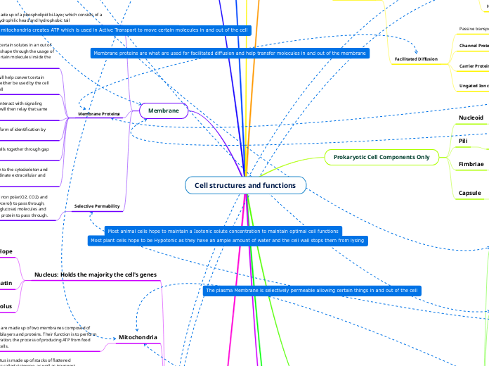
Made up of a phospholipid bi-layer, which consists of a hydrophilic head and hydrophobic tail
Depending on the phase transition temperature the membrane can either be fluid (where stuff can pass through easily) or rigid (where it is much more difficult for certain things to pass through). Unsaturated hydrocarbon tails = fluid while saturated hydrocarbon tails = rigid
Membrane Proteins^
Certain proteins help to transport certain solutes in an out of the cell, while others even change shape through the usage of ATP to allow only the transfer of certain molecules inside the cell.
Some proteins are enzymes that will help convert certain molecules so that they are able to either be used by the cell itself or possibly another type of cell
Some proteins are receptors that interact with signaling molecules such as hormones that will then relay that same information to the rest of the cell.
Some proteins are also used as a form of identification by other cells.
Certain proteins are used to join cells together through gap junctions or tight junctions
Certain proteins are used to attach to the cytoskeleton and ECM allowing the proteins to coordinate extracellular and intercellular changes
Selective Permability
Membrane only allows certain small non polar(O2, CO2) and uncharged polar molecules(H2O, glycerol) to pass through, large uncharged polar(Sucrose and glucose) molecules and Ions (Na+ K+) require the usage of a protein to pass through.
Non Polar
Any R group ending in CH or a 6 ringed carbon hexagon
Polar
Any R group ending in OH, SH, O, or NH2
Acidic
Any R group ending in o- causing the amino acid to have an overall negative charge
Basic
Any R group ending in a NH+, NH2+ Or NH3+ causing the overall charge of the amino acid to be positive
Active Transport
Movement from a low concentration to a high one through the usage of energy
Sodium-Potassium Pump
Starts off by bringing Na+ in then once a large amount is inside it starts to release Na+, and for every three Na+ it brings out 2 K+ molecules come in, going against the concentration gradient
Proton Pump
Uses ATP to transfer H+ from a low concentration gradient to a high concentration gradient
Cotransport
When the transport of a solute from a low to high concentration also indirectly transfers another substance
Gated Ion Channels
Stretch Gated
Ion gate opens only when membrane is deformed and will open up as a response to the stimuli
Ligand gated
Ion gate opens up in response to when a Neurotransmitter binds to the channel
Voltage Gated
Ion channel opens or closes depending on the membrane potential
Passive Transport^
Diffusion across the cell membrane with no energy usage
Diffusion
Movement of certain molecules from a high to low concentration until they reach an equilibrium
Osmosis
The diffusion of water from a concentration that has a low concentration of solute to the side that has a high concentration of solute until an equilibrium is reached
Water Balance
Isotonic
Solute concentration is the same inside and outside of cell so no water movement
Hypotonic
Cell has a greater solute concentration that outside so the cell starts to gain water
Hypertonic
The cell has a smaller solute concentration than outside so the cell starts to lose water
Facilitated Diffusion
Passive transport through the usage of proteins
Channel Protein
Channel proteins have a channel that allow solutes or other water molecules to pass through
Carrier Protein
Carrier proteins open up to intake certain molecules and then open back up to release them (pac-man)
Ungated Ion channel
Ion channel that lets ions just flow in and out of the cell from High to low concentrations
Nucleoid
The nucleoid is the region of cytoplasm in prokaryotic cells that holds the chromosome with most or all of the cell's genetic material.
Pili
The pili are structures on the surface of prokaryotic cells that are short and hair-like. Their role is to mate with other cells.
Fimbriae
Hairlike appendages that are typically shorter and more numerous than pili, and are used as a means for prokaryotes to stick to their substrate or to each other.
Capsule
The capsule is a sticky outermost layer that is made of polysaccharides or proteins and exists to help bacteria stick to organisms or surfaces. It also protects the bacteria from phagocytosis and dehydration.
Cell Wall
In plants and fungi (eukaryotes), the cell wall is made up of cellulose or chitin. In bacteria, it is made of the polymer peptidoglycan, but archaeal cell walls do not contain peptidoglycan, they only contain proteins and other polysaccharides. No matter what it is made of, the cell wall has the same purpose in every cell which is to provide structure and support, as well as to act as a semi-permeable surface to allow molecules to enter and exit the cell.
Plasma Membrane
In both prokaryotic and eukaryotic cells, the plasma membrane is made of a phospholipid bilayer embedded with proteins, but the lipids are negatively charged in prokaryotic plasma membranes while the lipids in eukaryotic plasma membranes are neutral. This difference is due to the fact that the plasma membrane in prokaryotes carries out all the functions that are done by the plasma membrane and the membrane-bound organelles of eukaryotes. Functions that are similar between the two are separating the cytoplasm from the cell's outside environment and controlling the passage of molecules that go in and out of the cell.
Cytoplasm
The cytoplasm is a gel-like fluid that is enclosed by the plasma membrane and is separate from the nucleus of eukaryotic cells. It functions as a medium for chemical reactions to take place inside both prokaryotic and eukaryotic cells.
Genetic Material
The genetic material (DNA) floats freely throughout prokaryotic cells which only have a singular chromosome in the nucleoid, while the DNA in eukaryotic cells is enclosed in a membrane-bound nucleus which contains multiple chromosomes. DNA itself is composed of building blocks called nucleotides, which are composed of a phosphate group, a deoxyribose sugar and a nitrogenous base. The role of DNA is storing information necessary to cell replication, mutation and gene expression.
Ribosomes
Ribosomes in prokaryotes are smaller than those in eukaryotes (70S vs 80S). Prokaryotic ribosomes are made of a 50S large subunit and 30S small subunit, while eukaryotic ribosomes are made of a 60S large subunit and 40S small subunit. The large subunit in prokaryotic ribosomes is made up of two rRNA molecules, 23S rRNA and 5S rRNA, while the large subunit in eukaryotic ribosomes is made up of three rRNA molecules, 28S rRNA, 5.3S rRNA and 5S rRNA. Both prokaryotic and eukaryotic ribosomes are composed of rRNA and ribosomal proteins, but prokaryotic ribosomes are 60% rRNA and 40% ribosomal proteins while eukaryotic ribosomes are 40% rRNA and 60% ribosomal proteins. Ribosomes in prokaryotic cells are scattered freely throughout the cytoplasm and even though eukaryotic cells also contain free ribosomes, most of their ribosomes are attached to the outer surface of their nucleus and endoplasmic reticulum. In both prokaryotic and eukaryotic cells, the ribosomes are the site of protein synthesis.
Flagella
Flagella are structures that can be scattered throughout the surface of cells or collected at one or both ends of the cell. In both prokaryotes and eukaryotes, flagella are made of proteins, but the proteins that make them up are completely different from each other in prokaryotes and eukaryotes. Additionally, flagella in bacteria and archaea are also made of completely different proteins. Flagella in prokaryotes are about a tenth of the width of flagella in eukaryotes. However, despite their structural differences, they all serve a common purpose which is cell mobility.
Structure
Phospholipids are composed of a hydrophilic (polar) phosphate head and two hydrophobic (nonpolar) hydrocarbon tails connected by glycerol.
Function
The structure of phospholipids allows them to form bilayers where their hydrophilic heads face outside and their hydrophobic heads face inwards to avoid water. These phospholipid bilayers are what make up the plasma membrane of cells, and they are mostly held together by hydrophobic interactions. Phospholipids are essential to membrane fluidity in that membrane fluidity will depend on the properties of the phospholipids. If the phospholipids have mostly unsaturated hydrocarbon tails, the kinks in the tails caused by their double bonds will prevent the phospholipids from being able to pack together as closely as phospholipids with saturated hydrocarbon tails, increasing membrane fluidity. Other factors that affect membrane fluidity are temperature and cholesterol. Lower temperatures decrease membrane fluidity unless cholesterol is present. If cholesterol is present, membrane fluidity will increase at lower temperatures and decrease at higher temperatures. This is because cholesterol limits phospholipids from becoming closely packed together which prevents the solidification and retains the fluidity of the membrane. In the case of higher temperatures, cholesterol restricts phospholipid movement and therefore decreases membrane fluidity.
Nucleus: Holds the majority the cell's genes
Nuclear Envelope
The nuclear envelope encloses the nucleus and separates it from the cytoplasm. It is a double membrane, with each membrane consisting of a lipid bilayer and proteins. This envelope also has pores that are lined by protein structures called pore complexes that control the entrance and exit of macromolecules, proteins and RNA.
Chromatin
Chromatin is a material made of DNA and proteins. It is the material that makes up a cell's chromosomes.
Nucleolus
The nucleolus is a nonmembranous structure composed of DNA, RNA and proteins. It works to produce the cell's ribosomes.
Mitochondria
Mitochondria are made up of two membranes composed of phospholipid bilayers and proteins. Their function is to perform cellular respiration, the process of producing ATP from food brought into cells.
Golgi Apparatus
The Golgi apparatus is made up of stacks of flattened membranous sacs called cisternae, as well as transport vesicles that travel between the Golgi and other organelles. The cisternae in the Golgi are not physically connected like they are in the ER. Golgi stacks have two different sides, a cis face that is typically located closer to the ER and a trans face where vesicles are released to travel off to different sites. The role of the Golgi apparatus is to modify materials it receives from the ER and sent the materials to other organelles or out into the cytoplasm.
Endoplasmic Reticulum (ER): A network
of membranous tubules and sacs called
cisternae that is continuous with the nuclear envelope. The ER is separated into two regions: the smooth ER and the rough ER.
Smooth
The smooth ER region is called this way because it does not
contain ribosomes. It has many functions in metabolic processes, such as using its enzymes to synthesize lipids and to detoxify drugs and poisons, as well as storing calcium ions.
Rough
The rough ER region gets its name from the fact that it is covered in ribosomes. It secretes proteins that are bonded to carbohydrates, structures which are called glycoproteins. It also uses its enzymes to create membrane phospholipids, and it adds membrane proteins and phospholipids to its own membrane in order to grow. Its membrane is held together by hydrophobic sections.
Lysosomes
Lysosomes are membranous sacs of hydrolytic enzymes.
The enzymes themselves are nearly neutral, but lysosomes are acidic in order to aid in intracellular digestion by using its enzymes to digest food and produce nutrients for the cell such as sugars and amino acids.
Chloroplasts
Similar to mitochondrion, chloroplasts are also made of two
membranes as well as internal membranous sacs, but they only appear in plants. These internal membranous sacs are called thylakoids, and some of these thylakoids appear in stacks called grana. The stroma is the fluid outside of the thylakoids, and it contains enzymes, ribosomes and the DNA of the chloroplast. All of these components of chloroplasts function together to perform photosynthesis.
Cytoskeleton: A network of fibers extending throughout the cytoplasm
Microtubules
Microtubules are larger in diameter than both microfilaments and intermediate filaments. They are hollow rods made of a globular protein called tubulin. Tubulin is a dimer consisting of the alpha and beta tubulin polypeptides. The microtubules provide the cell with shape and support, in addition to acting as a track where organelles with motor proteins can move throughout. They also play a role in separating chromosomes during cell division.
Centrosome
The centrosome is a region located near the nucleus, and it is the main site of microtubule assembly.
Intermediate filaments
Intermediate filaments are wider in diameter than microfilaments but smaller than microtubules. They are made of fibrous proteins coiled into cables, and their function is in maintaining the cell's shape, anchoring the nucleus and other organelles, as well as forming nuclear lamina.
Microfilaments
Microfilaments are smaller than both microtubules and intermediate filaments. They are made up of two intertwined strands of actin, and they work in maintaining cell shape, contracting muscles, changing cell shape, aiding in cell motility as well as cytoplasmic streaming in plant cells and cell division in animal cells.
Plasmodesmata
The plasmodesmata are only found in plants; they are membrane-lined channels filled with cytosol that connect cells into a continuous structure where water and small solutes can travel freely throughout, as well as some proteins and RNA molecules.
Peroxisomes
Vacuoles: large vesicles derived from
the ER and Golgi
Animal Cell Vacuoles
Vacuoles in animal cells are small, membrane enclosed
structures that function with lysosomes to digest food brought into the cell.
Plant Cell Central Vacuole
Mature plant cells typically have one large central vacuole
which absorbs water to support the growth of the cell and contains a solution called cell sap which acts as a receptacle for inorganic ions like potassium and chloride.
Animal Cell Junctions: All are composed
of epithelial tissue
Gap Junctions
Gap junctions are made of membrane proteins that create
space for small molecules to pass, and they are crucial to cell communication.
Tight Junctions
Tight junctions use proteins to bind plasma membranes of cells to each other so no fluids can pass between the cells.
Desmosomes
Desmosomes attach cells to each other sturdily and are held in the cytoplasm by intermediate filaments made of keratin proteins.
takes place in the chloroplast
includes
light dependent reactions
uses
light energy to make two molecules
ATP
the energy storage molecule
NADPH
the reduced electron carrier
takes place
thylakoid membranes
of organelles called
chloroplast
photosystems
Large protein and pigment complexes that are specially designed to gather light are essential for the light reactions.
two types of photosystems
photosystem 1
The electron arrives at photosystem I and joins the P700 pair of chlorophylls in the reaction center. The electron in P700 is then raised to an extremely high energy level and transmitted to an acceptor molecule when light energy is absorbed by pigments and sent inward to the reaction site.
Photosystem 2
Energy is transferred from pigment to pigment inside of photosystem II when light is absorbed by one of the numerous pigments inside. This process continues until the reaction center is reached. An electron's energy is raised there, where it is transmitted to P680. An acceptor molecule receives the high-energy electron.
releases
O2
start in
ETC
NADPH formation
the high energy electron is transferred through ETC and then at the end of the chain the electron is passed to NADP+ to make NADPH
pumps H+ ions into thylakoid
H+ then activates
ATP synthase
As it moves through an electron transport chain, the high-energy electron expends energy. A gradient is created when some of the energy released causes the pumping of ions from the stroma into the interior of the thylakoid.
then
H+ ions flow down their gradient and into the stroma, they pass through ATP synthase, driving ATP production
a process known as
Chemiosmosis
calvin cycle
where sugar is synthesized
takes place
Stroma
Cells and tissues that support and give structure to organs, glands, or other tissues in the body.
Phase 1
carbon fixation
adds CO2 from the atmosphere to ribulose bisphosphate using and enzyme Rubisco
this forms
6 carbon unstable intermediate
splits to form 2 molecules of
3-phosphoglyceric acid (3-PGA)
This reaction is catalyzed by the enzyme RuBP carboxylase/oxygenase, or rubisco
3-PGA molecules
phase 2
reduction
3-PGA molecules
uses
ATP
NADPH
to convert 3-PGA molecules into molecules of
G3P
produces
NADP+
Phosphate
phase 3
regeneration of CO2 acceptor
Some G3P molecules go to make glucose, while others must be recycled to regenerate the RuBP acceptor.
requires
ATP
and involves
complex network of reactions
A process where plants use sunlight to make food from water and carbon dioxide
Glycolysis: First stage of cellular respiration (takes place in the cytosol)
Key Steps
Step 1: Hexokinase transfers a phosphate group from ATP to glucose, making it more chemically reactive. The charge on the phosphate traps the sugar in the cell.
Step 3: Phosphofructokinase transfers a phosphate group from ATP to the opposite end of the sugar, using a second molecule of ATP.
Input
1 Glucose
2 NAD+
2 ATP
Output
2 Pyruvate
2 NADH
4 ATP
Formed through substrate level phosphorylation
Net
2 Pyruvate
2 NADH
2 ATP
Pyruvate Oxidation (starts in the cytosol and moves to the mitochondrial matrix)
Input
2 Pyruvate
2 CoA
2 NAD+
Output
2 Acetyl CoA
2 NADH
Net
2 Acetyl CoA
2 NADH
Krebs Cycle/Citric Acid Cycle (takes place in the mitochondrial matrix)
Key Steps
Step 1: Acetyl CoA adds its two-carbon group to oxaloacetate, forming citrate.
Step 3: Isocitrate forms alpha ketoglutarate through an oxidation-reduction reaction.
Input
2 Acetyl CoA
6 NAD+
2 FAD
Output
6 NADH
2 FADH2
2 ATP
Formed through substrate level phosphorylation
Net
6 NADH
2 FADH2
2 ATP
Oxidative Phosphorylation
Electron Transport Chain (ETC))
The electron transport chain is a series of molecules found in the inner membrane of the mitochondria in eukaryotes, and found in the plasma membrane of prokaryotes. Most of the chain consists of proteins with electron carriers that exist in multiprotein complexes numbered I through IV. In this chain, electrons from either NADH or FADH2 move down the chain through increasingly electronegative carriers in a series of oxidation-reduction reactions. The electrons are transferred to oxygen at the end of the chain, and this oxygen forms water by splitting in half and taking up H+. The transport of electrons and pumping of protons create an H+ gradient across the membrane. This whole process releases an abundance of free energy which is coupled with chemiosmosis to synthesize ATP.
Chemiosmosis
The free energy released from the ETC is used to pump H+ ions from the mitochondrial matrix to the intermembrane space, creating an electrochemical gradient. The flow of H+ ions down this gradient and back to the mitochondrial matrix synthesizes ATP due to the passing of the H+ ions through the enzyme ATP synthase. When H+ ions flow down their electrochemical gradient, ATP synthase turns like a waterwheel, catalyzing the addition of an inorganic phosphate to ADP in order to form ATP.
Input
O2
10 NADH
2 FADH2
Output
H2O
26-28 ATP
Formed through the coupling of chemiosmosis with the ETC
Net
H2O
26-28 ATP
Local signalling
Local
Paracine Signalling
Secreting cell releases ligand/a signal to target cell receptor in its local area
Synaptic Signalling
Electrical signal along nerve cell causes the release of neurotransmitters that diffuses across synapses into target cell receptors
A stimulus that is strong enough to cause depolarization will result in triggering an action potential
Long distance
If the target cell is too far away a cell may use a medium like a blood vessel to transport signals like hormones across the body to target cells receptors
Physical Contact
Cells communicate through physical touch through the use of junctions allowing molecules to transfer or proteins to bind
Gap Junctions
Gap Junctions are in animal cells
Plasmodesmata
Plasmodesmata are in plant cels
Receptor
Intracelluar Receptors
Used with small non polar molecules and hormones that can easily bypass the membrane and bind to the receptor inside the cell
steps
Hormone/nonpolar molecule passes through the plasma membrane
The hormone/nonpolar molecule then binds to the receptor inside the cell activating it and becoming a receptor complex inside the cytoplasm
The receptor complex then enters the nucleus and binds to specifics genes
The bound proteins is then used as a transcription factor transcribing the gene into mRNA
Finally, the mRNA is translated into a specific protein
Membrane Recetpors
Used with larger hydrophilic polar molecules that can't enter the inside of the cell so they bind to the receptor on the outside of the cell.
steps
Reception
Transudction
Response
G protein
steps
G protein is bound to GDP making it inactive
Signalling mollecule binds to GPCR which allows for G protein to bind to it and caused GDP to be replaced with GTP
The now activated G protein can now travel and activate a nearby Enzyme triggering a cellular response
G protein then removes phosphate group from GTP making it once again GDP deactivating the G protein
Tyrosine Kinase
kinase help remove phosphate groups from ATP and add them to proteins
steps
Made up of two polypeptides that when a signal molecule is added to each of them dimerize
Each polypeptide functions as a kinase and adds phosphate groups from ATP to the other polypeptide (autophosphorylation)
When a total of 6 phosphate groups have been added with three on each polypeptide the tyrosine-kinase if fully activated
Now the receptor can bind to other proteins to create a cellular response in the cel
Phosphorylation Cascade
a type of signal transduction pathway that uses kinases and phosphates.
A signal molecule attaches to a receptor which in turns activates a relay molecule
The relay molecule activates the first protein kinase
The protein kinase then activates a different kinase
The first kinase has its prostate groups removed by a phosphatase
And then this process repeats until a kinase adds a phosphate group to a protein activating it and bringing a cellular response
The cellular response can either be the synthesis of enzyme or other proteins, by turning genes off and on
Second Messengers
Second Messengers are relay molecules which carry the message from the first signal inside the cell
cAMP
steps
A type of second messenger Synthesized from ATP using Adenylyl Cyclase^
cAMP binds to another kinase activating it
cAMP is then converted to AMP by Phosphodiesterase
Ion channell Receptor
An ion channel receptor acts as en entrance for ions.
steps
An ion channel remains closed until a signaling molecule binds to it
When the ion channel opens up only certain molecules are allowed through such as Na+ or Ca+
Finally when the signaling molecule leaves the ion channel closes along with it
Alcohol Fermentation
Process
Carbon dioxide is released from pyruvate which is converted into acetaldehyde. The acetaldehyde is then reduced by NADH to ethanol, regenerating the NAD+ necessary for glycolysis to keep occurring.
Input
2 NADH
2 Pyruvate
Output
Ethanol
NAD+
Lactic Acid Fermentation
Process
Pyruvate is directly reduced by NADH to form lactate with no release of carbon dioxide involved.
Input
2 NADH
2 Pyruvate
Output
Lactate
NAD+
involves
Ribosomes
consist of
rRNA
has
3 active sites
A-site
P-site
growing polypeptide
E-site
exit site
small ribosome
large ribosome
protein synthesis
starts on
Free Ribosomes
enter
endomembrane system
a collection of organelles and membranes in eukaryotic cells that collaborate to transport, package, and alter lipids and proteins.
with the help of
SRP
(signal recognition particle and is made of RNA and protein)
then proteins are escorted to the
Rough ER
Here, several carbohydrate groups are added by enzymes in the ER to this protein now its called a glycoprotein
From the ER, the protein is shipped through a vesicle to the
Golgi
Proteins and lipids go through further changes as they go through the Golgi.
then the proteins can be transported to the
Plasma membrane
stay as
Membrane protein
be secreted out of the cell
Lysosome
synthesis completes on free ribosomes which lead proteins to organelles like :
Mitocondria
Peroxisomes
cytoplasm
nucleus
Plastids
Structure
A polymer made up of monomers called nucleotides which are connected through phosphodiester bonds. RNA is also composed of nucleotides linked by phosphodiester bonds.
Replication
It was determined by Messleson and Stahl’s experiment that DNA replicates semiconservatively, meaning that the daughter molecules of a parent DNA molecule will each have one strand from the parent molecule and one newly made strand.
Prokaryotes
Site of transcription: Cytoplasm
Initiation
RNA polymerase binds to the template strand’s promoter sequence.
Elongation
RNA polymerase moves along the template strand of DNA, adding ribonucleotides in the 3’ to 5’ direction.
Termination
RNA polymerase reaches the template strand’s terminator sequence, stopping transcription and releasing mRNA that needs no additional alterations unlike in eukaryotes.
Eukaryotes
Site of transcription: Nucleus
Initiation
Proteins called transcription factors bind to the template strand’s promoter sequence, and RNA polymerase II binds the promoter.
Elongation
RNA polymerase II moves along the template strand of DNA , adding ribonucleotides to the 3’ end of the growing strand of RNA.
Termination
RNA polymerase II reaches the terminator sequence, stopping transcription and releasing pre-mRNA.
RNA processing
The pre-mRNA ‘s 5’ end gets a cap and its 3’ end gets a polyA tail which is added by polyA polymerase. Then spliceosomes are used to remove the pre-mRNA’s introns and splice together its exons, forming mRNA.
Eukaryotes
Most gene regulation occurs at the transcription level as there is no need to from pre mRNA if no protein is needed
Transcription factors
Proteins that bind to the promoter and are crtical for gene expression and regulation
Specific
Transcription factors that bind to distal control elements which are called enhancers, which can be either close to t
Activators
proteins that comes from cell signaling which increases levels of transcription
DNA Bending protein
brings the bound activators closer to the promoter, which results in the activator binding to certain mediator proteins forming an active transcription complex on the promoter
Repressors
Reduce levels of transciption
General
Transcription factors that bind to the proximal control elements on or near the promoter, resulting in basal/background level of transcription
Prokaryotes
Like Eukaryotes most gene regulation occurs at the transcription level
Operons
A segment of DNA positioned within the promoter that turns on gene expression or turns it off
Positive Regulation
Whenever an Acitvator binds to a Operator resulting in turning the gene ON and creating a high level of gene expression
Negative Regulation
When a repressor binds to a Operator causing the gene expression to be OFF (or basal).
Lactose
when lactose is used as an energy source Permease, a protein, will transfer the lactose while B-Galactosidase will break the glycolosidic linkage forming glucose an galactose
Lac Operon
When Lactose is present the transcription of LacZ, LacY, and LacA is blocked, so the repressor is able to bind to the operator and the operon is OFF
When glucose is present the lac operson is off due to glucose blocking Adenyl Cyclase
When lactose is present the repressors binds to it instead of the operator resulting in RNAP being able to bind to the promoter, through the activator protein CAP which is activated through cAMP that is formed from adenyl cyclase-Operon ON
When ON the mRNA is transcribed and translated to form 3 proteins, B-Galactosidase, Permease, and Transacetylase which all help break down lactase
Prokaryotes
Occurs at the same time as transcription
Eukaryotes
Occurs at different times compared to transcription
tRNA
tRNA going in the 3-5 directions binds to mRNA going in the 5-3 direction through the use of an anti codon and binds to the small ribosomal unit
Aminoacyl tRNA
add amino acid to tRNA
Large ribosomal unit
The start codon binds to the anticdodon in the p site, another trna then binds to the mRNA in the A site, then the p site trna moves to the e site and exits the ribosomal complex after losing its amino acid
Pepitdeyl Transferase
Add polpeptide bonds to amino acids to join them together
termination
When the stop codon reaches the a site a realease factor helps the ribsomola complex break apart and stops translation
Nucleosome
DNA wraps around a histome core twice forming a Nuclesome
Histome Core
A histome core is composed of multiple proteins which consist of H2A, H2B, H3, H4 with the H1 being on the outside of the core
H1
The H1 protein help the DNA wind even tighther together and eventually this winding results in the formation of a chromosome