Membrane Bound Organelles
Nuclear envelope- double membrane enclosing the nucleolus
Lined in nuclear Lamina-
A netlike array of protein filaments that lines the inner surface of the nuclear envelope and helps maintain the shape of the nucleolus
Cytoplasm- surrounds the organelles
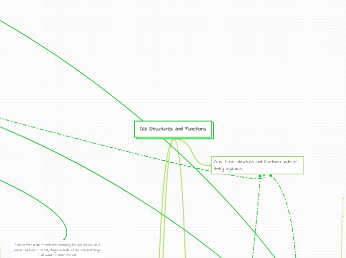
Plasma Membrane-
membrane enclosing the cell; serves as a barrier between the cell, things outside of the cell, and things that want to enter the cell
Consists of double layer of phospholipids with various proteins attached to or embedded in it
Hydrophobic parts are found in the interior of membrane
Hydrophilic regions are in contact with aqueous solutions on either side
Carbohydrate side chains attached to proteins on the outside of the cell that are embedded into the plasma membrane
Membrane Transport and Structure
Phospholipid Bilayer
Amphipathic
contains both hydrophobic and hydrophilic components
Phosphate Group head
and Lipid tail, connected by Glycerol
Presence of Cholesterol
Membrane Fluidity
Above Temp: crystalline phase, fluid, unsaturated hydrocarbons tails
Below Temp: gel phase, rigid, saturated hydrocarbon tails
Membrane Proteins
Peripheral Proteins
- proteins located on the outside
of the plasma membrane
Transmembrane (Integral) Proteins
- proteins that are located within the plasma
membrane
- composed of alpha helices, N-terminus outside and C-Terminus inside cell
Different Functions of Membrane Proteins
Transport Proteins
and Membrane Transport
Selective Permeability
- regulates cell movement, cell traffic, and inter/extracellular interactions
HIGH: small, nonpolar
MEDIUM:-HIGH: small, uncharged polar
MEDIUM-LOW: large, uncharged polar
LOW: ions
Passive Transport
- Type of transport that doesn't require ATP
- High to Low
- Down Concentration Gradient
Diffusion
- the movement of particles down the concentration gradient
Facilitated Diffusion
- The diffusion of water and other polar molecules with the help of a protein that has a hydrophilic interior channel (aquaporin)
Osomosis
- the movement of water down the concentration gradient from high to low to help establish equilibrium, or water balance
Isotonic:
- solute concentration same inside and outside the cell
- no net water movement
ANIMALS: Normal
PLANTS: Flaccid
Hypotonic:
- the solute concentration less than inside the cell
- water moves inside the cell
ANIMALS: Lysed
PLANTS: Turgid (Normal)
Hypertonic:
- the solute concentration greater than inside the cell
- water moves outside the cell
Singled-Celled eukaryotes have contractile vacuoles that push out unnecessary water
Active Transport
- Type of transport that REQUIRES ATP
- Low to High
- Against Concentration Gradient
Pumps
Sodium Potassium Pumps
- Ions bind to the proteins stimulating phosphorylation by a kinase using ATP
- 3 Na+ transported outside, 2 K+ transported inside
Proton Pumps
Electrogenic Groups
- Transmembrane protein that generates voltage across the membrane establishing membrane potential
- Helps store energy for cellular work
CoTransport
- The transport of multiple substances simultaneously
- Driven by concentration gradient
- EX: sucrose moving into the cell as H+ diffuses out
Bulk Transport
- Vesicles and ATP are used to move fluids, solutes, and molecules in and out of the cell
EXocytosis:
- EXiting a cell:
ENdocytosis:
ENtering a cell
Phagocytosis:
Large food particles enter the cell
Pinocytosis:
Dissolved fluids enter the cell
Receptor-Mediated Endocytosis:
Receptors deployed by the cells bind to specific particles and bring them to the cell
Enzymatic Activity
- Carries out metabolic processes
- Membranes have active sites and chemical receptors that carry out sequential steps
Signal Transduction
- Possess certain receptors that are fit to receive messages from signaling molecules
- Molecules binds to cytoplasmic proteins which have ends that are outside and inside the cell.
Cell-Cell Recognition
- Glycoproteins serve as ID tags which are recognized by certain proteins and receptors of membrane cells
- Short-Lived Connection
Intercellular Joining
and Cell Junctions
- Membranes cells hook together in junctions
- Long-Lived Connection
Animal Cells
Gap Junctions:
Intercellular transport and cell signalling/communication
Proteins Present:
connexin proteins
such as kinases
Tight Junctions:
these junctions form continuous seals with the help of a protein that keeps solutes and fluids from moving across cells
Proteins Present:
occludin, claudin, and accessory proteins
Desmosomes:
similar to tight junctions but not as tight allowing selective fluids and solutes to pass through
Proteins Present:
2 cadherins, DSG2, DSC2, and 3 cytoplasmic plaque proteins
Plant Cells
Plasmodesmata:
- connects membranes and cytoplasm of adjacent cells- intercellular transport and cell signaling/communication
Attachment to ECM and Cytoskeleton
Connects to the cytoskeleton's
microfilaments non-covalently
The connection to the ECM helps
cell perform inter/extracellular changes
Endoplasmic reticulum-network of membranous sacs and tubes; active in membrane synthesis and other
Rough ER-
ribosomes are attached
secretes proteins
transport vesicles move the protiens
membrane factory for the cell
Smooth ER
synthesis of lipids, metabolism of carbohydrates, detoxification of drugs and poisons, and storage of calcium ions
steroids are produced
Golgi apparatus- organelle active in synthesis. modification, sorting, and secretion of cell products
products of the ER are modified and stored then sent to other destinations
consists of flat membranous sacs, called cisternae, stacked together
cis face, same side, is located near ER
trans face, opposite side is located gives rise to vesicles that pinch off and travel to other sides
vesicles around the Golgi engage in the transfer of material
Lysosomes-digestive organelle where macromolecules are hydrolyzed
carry out intracellular digestion in a variety of circumstances
Cytoskeleton- reenforces cell shape; functions in cell movement; components are made of proteins
cell motility-changes in cell location and more limited movement of cell parts
motor proteins-
interacts with cytoskeletal elements and other cell components, producing movement of the whole cell or parts of the cell
Structures and functions of Cytoskeleton
Microtubules
maintenance of cell shape, cell motility, chromosome movements in cell division; organelle movements
involved in the separation of chromosomes during cell division
grow out of centrosomes
Hollow rods constructed from a globular protein called tubulin, each tubulin protein is a dimer
made up of alpha-tubulin and beta-tubulin
can be disassembled and used to build microtubes somewhere else
grow in length by adding tubulin dimers
Microfilaments
Maintenance of cell shape; changes in cell shape; muscle contraction; cytoplasmic streaming (plant cells); cell motility, cell division (animal cells)^
form structural networks when certain proteins bind along the side of such a filament and allow a new filament to extend as a branch
strutural role to bear tension
thin solid rods
also called actin filaments because that are built from actintwisted chain double chain of actin subunits
use a motor proteins called myosin to cause contraction of muscle cells
Intermediate filaments
maintenance of cell shape; anchorage of nucleus and certain other organelles; formation of nuclear lamina
more permanent fixtures of cells, often disassembled and reassembled in various parts of cell
Mitochondrion- cellular respiration occurs and ATP is generated
Produces ATP which is energy that the cell uses
outer membrane is smooth, but the inner membrane is convoluted, with folding called cristae; there are two compartments
intermembrane space- between the inner and outer membranes
mitochondrial matrix-
compartment of the mitochondrion containing enzymes and substrates for the citric acid cycle, as well as ribosomes and DNA
Cellular Respiration
1. Glycolysis
(Steps 1 and 3)
- occurs in the cytoplasm
- Energy Investment and Energy Pay Off Phases
- ATP formed with Substrate-Level Phosphorylation (sLP)
INPUTS
- 1 Glucose
- 2 NAD+
- 2 ATP
OUTPUTS
- 2 Pyruvate
- 2 NADH
- 4 ATP
2. Pyruvate Oxidation
- occurs from the cytosol to the Mitochondrial Matrix
INPUTS
- 2 Pyruvate
- 2 CoA
- 2NAD+
OUTPUTS
- 2 Acetyl CoA
- 2 NADH
3. Citric Acid Cycle
(steps 1 and 3)
- occurs in the Mitochondrial Matrix
- ATP formed with SLP
INPUTS
- 2 Acetyl CoA
- 6 NAD+
- 2 FAD
OUTPUTS
- 2 ATP
- 6 NADH
- 2 FADH2
4. Oxidative Phosphorylation
- occurs among the Mitochondrial Matrix, Inner Mitochondrial Membrane, and the Inner-membrane Space
Electron Transport Chain
- NADH: I, Q, III, Cyt C, and IV
- FADH2: II, Q, III, Cyt C, and IV
- Oxygen is the final electron acceptor
As electrons move across the complexes, H+ is pumped out into the intermembrane space establishing a proton gradient.
Chemiosmosis
The proton gradient coming back into the organelle through ATP Synthase produces ATP. Gradient spins motor that attaches the inorganic phosphate to ADP to create ATP.
The ETC and Chemiosmosis are coupled to generate ATP. The gradient from ETC is required to produce ATP with ATP Synthase.
INPUTS
- O2
- 10 NADH
- 2 FADH2
OUTPUTS
- H2O
- 26-28 ATP
Microvilli- increase the cells surface area
Peroxisome-
organelle with various specialized metabolic functions; produces hydrogen peroxide as a by-product and then converts it to water
Flagellum-
motility structure present in some animal cells, composed of a cluster of microtubules within an extension of the plasma membrane
Centrosomes-
region where the cell's microtubules are initiated; contains a pair of centrioles
p
use DNA to make proteins
carry out protein synthesis
consists of a small and large subunit
build proteins in two cytoplasmic locales
Free ribosomes-
suspended in the cytosol
Bound ribosomes-
attached to the outside of the endoplasmic reticulum or nuclear envelope
proteins destined for insertion into membranes; packing certain organelles suck as lysosomes or for export from the cell
Endomembrane system-
membranes of this system are related either through direct physical continuity of transfer by vesicles
Vesicles-
a membranous sac in the cytoplasm
Vacuoles-
large vesicles, that have many different function, selective in which solutes to transport
Extracellular Matrix (ECM)
Specific to animal cells as plants have cell walls
COMPONENTS
Collagen fibers embedded in a web of proteoglycan complexes
Proteoglycan Complexes:- hundreds of proteoglycan molecules attached non-covalently to a single long polysaccharide molecule
Fibronectin attaches the ECM to the integrins in the Plasma Membrane
- Integrins:: membrane proteins with two subunits
- Binds to the ECM on the outside and microfilaments on the inside
- Helps transmit signals
Carbohydrates
carbon skeletons (can vary in..)
length
branching
double bond position
presence of rings
Isomers-compounds that have the same
# of atoms of the same elements but
different structures and properties.
Structural Isomers (differing placement of
same atoms/elements)
Geometric Isomers
cis isomer: on the same side
trans isomer: on opposite sides
Enantiomers (mirror images: differ due to the presence of an asymmetric carbon bonded to 4 different groups)
Polymers
Synthesis of Polymers: condensation/dehydration reactions
Breakdown of POlymers: hydrolysis
Functional Groups-chemical groups that affect
function by being directly involved in chemical interactions.
Hydroxyl, Carbonyl, Carboxyl, Amino, Sulfhydryl, Phosphate, and Methyl group.
Carbohydrates serve as fuel and building material
Aldoses (CO group at the end)
Glucose
Alpha glucose=starch
Beta glucose=cellulose
Galactose
Ketoses (CO group on the inside)
Fructose
Monosaccharides
Polysaccharides: formed when 100
or more monosaccharides are bonded
together through glycosidic linkages.
Storage Polysaccharide (energy)
- glycogen (animals)
- starch (plants)
- dextran (bacteria)
Structure Polysaccharide
- cellulose (Beta glucose) (plant cell walls)
- chitin (exoskeleton of insects, cell walls of fungi)
Disaccharide Synthesis: Glucose + Fructose = Sucrose
(Glycosidic bond/linkage formed from dehydration synthesis)
Lipids
Fats
Triacylglycerol/triglyceride
1 glycerol & 3 fatty acids
C3H5O3 (glycerol)
Palmitic Acid (3 fatty acids)
Saturated
- animal sources
- solid at room temperature
- NO double bonds
Unsaturated
- plant sources
- liquid at room temperature
- double bonds
cis (hydrogen on the same side)
causes molecule to have a bend
trans (hydrogen on opposite sides
-called trans fat)
Phospholipids
make up the phospholipid bilayer (made out of hydrocarbons), which contain non polar covalent bonds= hydrophobic
Steroids
contain 4 fused rings
found in animals
common component in membranes
ex: cholesterol
Proteins
Primary Structure
- sequence of amino acids/polypeptide (main chain)
Covalent Bonds
Peptide Bond- the basic bond that links amino acids in all protein structures ( through dehydration synthesis)
Di-sulphide bond- formed by the coupling of two thiol (–SH) groups/the only covalent bond between R-groups (intramolecular)
Secondary Structure
- main chain turns and folds
- alpha helices (twists)
- beta pleated sheets (folds)
- intermolecular (between 2 or more structures)
Hydrogen Bond- interaction involving a hydrogen atom located between a pair of other atoms having a high affinity for electrons
Peptide Bond- the basic bond that links amino acids in all protein structures
Di-sulfide bond- formed by the coupling of two thiol (–SH) groups.
Tertiary Structure
- polypeptide folds into a 3D shape
through interaction of R-groups
Hydrophobic bonds- the tendency of nonpolar groups or molecules to aggregate in water solution.
Hydrogen Bonds- interaction involving a hydrogen atom located between a pair of other atoms having a high affinity for electrons
Ionic Bonds- type of linkage formed from the electrostatic attraction between oppositely charged ions in a chemical compound.
Van der Walls forces - the relatively weak attractive forces that act on neutral atoms and molecules and that arise because of the electric polarization induced in each of the particles by the presence of other particles.
Di-sulphide bonds- formed by the coupling of two thiol (–SH) groups.
R groups
covalent bonds
A covalent bond is formed when two atoms exchange one or more pairs of electrons.
ionic bonds
When an element takes an electron from another to finish an octet level, an ionic bond is created. relates to electro negative as well.
Acidic and Basic
Although acidic and basic groups are hydrophilic, they are joined by ionic bonds.
Ion dipole bonds
Electron-electron interactions between two neutral molecules with dipoles cause ion dipole forces.
between r-groups in an acid and water
nonpolar groups
CH
hydrophobic interactions will occur
polar groups
OH, NH, SH, CO
hydrogen bonds will form
Quaternary Structure
- tertiary chains coming together (structure
maintained by interchain interactions)
Hydrophobic bonds- the tendency of nonpolar groups or molecules to aggregate in water solution.
Hydrogen bonds- interaction involving a hydrogen atom located between a pair of other atoms having a high affinity for electrons
Ionic bonds- type of linkage formed from the electrostatic attraction between oppositely charged ions in a chemical compound.
Van der Walls forces- the relatively weak attractive forces that act on neutral atoms and molecules and that arise because of the electric polarization induced in each of the particles by the presence of other particles.
Di-sulphide bonds- formed by the coupling of two thiol (–SH) groups.
Nucleic Acids
DNA (deoxyribonucleic acid)
provides own information for replication
directs synthesis for mRNA and controls protein synthesis
gene expression
translation (info from mRNA makes proteins)
transcription (info from DNA is used to make RNA)
RNA (ribonucleic acid)
adenine, cytosine, uracil, and guanine
Organelles
Fimbriae- Bacteria can attach to particular receptor structures thanks to attachment structures.
Nucleiod- DNA situated here that is not membrane-enclosed
Cytoplasm- contains organic molecules and enzymes and is primarily composed of water.
Cell wall- rigid, external to the plasma membrane, provides security and shape
Glycocalyx- An outer layer is made up of a slime or capsule layer.
conserves fluids and guards the cell from being swallowed by other organisms. fights off phaagocytosis
Plasma Membrane- Selectively The cytoplasm is enclosed and controls the flow of substances during nutrition and waste transport as well as the location of metabolic processes.
Ribosomes-protein production and synthesization
Producers
Autotroph
Photoautotroph
Organism types:: Photo synthetic prokaryotes algae
Energy comes from a light carbon source CO2 HCO3
Chemoautotroph
Energy comes from in organic chemicals carbon source CO3 HCO3
Heterotroph
Photoautotroph
the sources are Light and organic compounds
Chemoautotroph
Organic Compounds
a nucleoside DOES NOT have a phosphate group
purines (A and G)
2 carbon nitrogen ring bases
pyrimidines (C, T, and U in RNA)
1 carbon nitrogen ring bases
joined by condensation/dehydration
reactions= phosphodiester bond/linkage
Functions
Caries out the transduction part of the signaling pathway
Most other intracellular receptors function in the same way
Details
1. Found in the cytoplasm or nucleus of target cell
2. Signaling molecule passes through target cell's plasma membrane
Steroid hormone interactions
1.The steroid hormone passes through the plasma membrane
2. Hormone binds to a receptor protein in the cytoplasm activating it
3. The hormone-receptor complex enters the nucleus and binds to specific genes
1.The hormone receptor complex turns on genes in a cells DNA function by being transcribed and processed into messenger RNA in the nucleus
2. Leaves the nucleus and is translated into specific proteins by ribosomes in the cytoplasm
3. Proteins called transcription factors control with genes are tuned on/ transcribed in mRNA
4. The bound proteins acts as a transcription factor , stimulating the transcription of the gene into mRNA
5. The mRNA is translated into a specific protein
G Protein linked Receptor
1. First messenger binds to GPCR, activating it
2. Activated GPCR binds to G protein, which is then bound by GTP, activating G protein
3. Activated G protein/GTP binds to adenylyl cyclase. GTP is hydrolyzed, activating adenylyl cyclase
4. Activated adenylyl cyclase converts ATP to cAMP
5. cAMP (second messenger) activates another protein, leading to cellular responses
Tyrosine Kinase Receptor
(made of 2 polypeptides
which dimerize when a
signal molecule is bound to
each polypeptide)
1. Signal molecule binds the each of the binding-sites
2. Polypeptides dimerize (join together), activating them (unphosphorylated dimer)
3. Autophosphorylation (phosphate group taken from ATP and added to the other polypeptide)
4. Fully activated receptor tyrosine-kinase (phosphorylated dimer)
5. Activated receptor now interacts with other relay proteins to bring out cellular response 1/2
Ion Channel Receptor
(acts as a gate for ions
when the receptor changes
shape)
gate allows specific ions (Na+ or Ca+) through a channel
very important in the nervous system
movement of ions in these channels may change voltage across membranes = triggers action potential
Action potential-
electrical signal that propagates along the membrane of a neuron or other excitable cells as a nongraded depolarization
COMPONENTS
m and tRNAs | Ribosomal Components:
E, P, A sites | large and small subunits | peptidyl transferase, amino-acyl tRNA synthetase | stop, start codons and anticodons | initiation, elongation, and release factors
PROCESS
:1. INITIATION:
- mRNA, tRNA, two ribosomal subunits come together
- GTP and initiation factors required to assemble this complex
- Small ribosome and tRNA bind to 5’cap, scans mRNA for AUG, and then large subunit assembles to form translation initiation complex
2. ELONGATION
- tRNA to A site with amino acid, moves to P site to for peptide bonds, then tRNA exits at E site
- peptide chain grows at the P site
- Each step GTP is used with GDP+P remaining
- Amino acids are added from N(amino) to C (carboxyl)
- peptidyl transferase creates the peptide bonds
- amino-acyl tRNA synthetase adds the appropriate amino acid after reading the mRNA codon
3. TERMINATION
- Stop codon read, release factors sits at A site, and then breaks apart the complex
- Driven by GTP
POST-TERMINATION
- Endomembrane System If it has an ER signal sequence, free ribosomes to the ER to the golgi body, out the cell/other locations via vesicles
- Amino acid signal sequence tell proteins where to go
- Secretory Pathway: path take protein from synthesis to modification to secretion
- Glycoproteins formed from glycosylation, (process of adding carbohydrate groups to proteins)
- Protein destinations: mitochondria, chloroplast, peroxisomes, nucleus
PROKARYOTES VS. EUKARYOTES
PROKARYOTES
- Occurs in the cytoplasm and uses mRNA and tRNA
- Ribosomal components: 70S, Large subunit: 50S (31 proteins) and Small subunit: 30S (21 proteins)
- All steps of translation the same, but elongation uses formal-MET, and lack of organelles means protein destinations are cytosol or plasma membrane
EUKARYOTES
- starts at free ribosomes (cytosol) but moves to
ER if initiated
- Ribosomal components: 80S, Large subunit: 60S (~ 45 proteins) and Small subunit: 40S (~ 33 proteins)
- these have more possibilities with more organelles and pathways than prokaryotes, and it just uses MET
Prokaryotes vs. Eukaryotes
Procces
Your text here
Your text here
Your text here
cytosol
Mitochondrion
Perixsome
Chloroplast
Nucleus
Endoplasmic Reticulam
Golgi Apparatus
cell exterior
plasma membrane
lysosomes
other parts of end-membrane system
In the cytosol, polypeptide synthesis starts on a free ribosome.
A signal peptide is bound by a signal recognition particle (SRP), the synthesis is momentarily stopped.
A receptor in the ER membrane is where SRP binds.
Polypeptide synthesis resumes when SRP leaves.
A receptor protein complex enzyme splits the signal peptide.
After leaving the ribosome, the finished polypeptide assumes its final configuration.
Becomes membrane protein
secreted outside the cell
Glycosylation
Glycoprotein (Carbohydrates and Protiens)
Secondary Protiens
Amylase is released by digestive enzymes.
Insulin is secreted by peptide hormones.
Casein is secreted by milk proteins.
Albumin is secreted by serum proteins
Proteins from extracellular matrix secrete collagen
additional locations