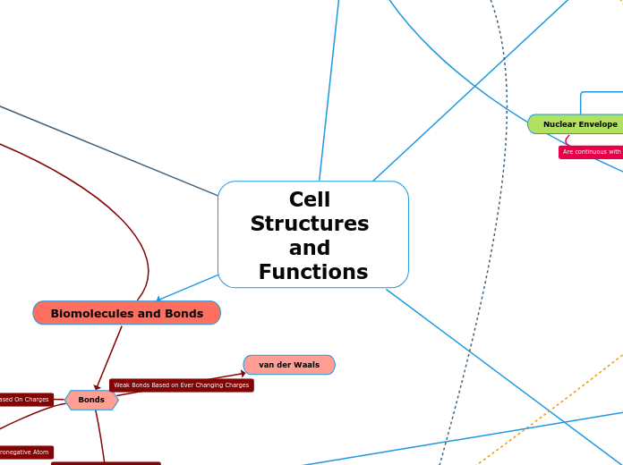
Bonds
Biomolecules
Carbohydrates
Extracellular matrix
Proteins
Mosaic Plasma Membrane
Proteins that do not move. Anchored to either the cytoskeleton or the ECM. Classified based on location.integral protein peripheral protein
Peripheral proteins
Anchored to the membrane.
Integral proteins
Inserted into the membrane either partially or fully. Fully inserted are called transmembrane proteinsSpans the entire membrane, part out and part inside
Lipids
Cholesterol
Affects fluidity and is an amphipathic molecule (hydrophobic and hydrophilic).presence between phospholipids reduces movement at low temp. prevents packing between phospholipids that otherwise would solidify the membrane.
Phospholipids
Has hydrophobic fatty acid tails and a hydrophilic head. (Amphipathic molecule) Have a specific phase transition temperature. Above this temp. lipid is in liquid crystalline phase and is fluidBelow this temp. lipid is in gel phase and is rigid. Mainly in charge of membrane fluidity - lets molecules pass through Phospholipid has more unsaturated fatty acids = more fluidUnsaturated fatty acids have one or more double bonds in the hydrocarbon chains of the fatty acidSaturated fatty acids have single bonds in the hydrocarbon chains of the fatty acid
Microfilaments
Muscle movement Actin and myosin cause movement Push and release (contraction) Amoeboid movement localized contraction/relaxationsqueeze interior fluid to cause movementCytoplasmic streaminglocalized contractions
Bacteria
Structures / Functions
Plasma Membrane
selectively permeable barrier
components - phospholipids, proteins
Nucleoid
DNA
plasmid - DNA seperate from the main
bacterial chromosome
Eukaryotes
Initiation
1. The small ribosomal subunit binds to the 5' end of the mRNA made in Transcription once it enters the cytoplasm.
2. The initiator tRNA pairs with the start codon on the mRNA.
3. The large ribosomal subunit attaches itself and the translation initiation complex is formed (this is due to the hydrolysis of GTP). When attached the tRNA is placed in the P site where the peptide chain can begin at the N-terminus with the amino acid MET.
Elongation
1. Codon Recognition - A charged tRNA
pairs it's anticodon with the mRNA codon
currently in the A site of the ribosome. Hydrolysis of GTP makes this process more accurate.
2. Peptide Bond Formation - The rRNA in
the large ribosomal subunit forms a peptide bond between the carboxyl end of the peptide chain and the new amino acid in A site. In doing this the amino acid and tRNA on P site disconnect and the tRNA is now empty.
3. Translocation - The empty tRNA moves to the E site and is released from the ribosome. The tRNA connected to the peptide chain is moved to the P site (the mRNA is shifted as well) and the A site is empty. The energy from GTP hydrolysis is used here. The cycle is then ready to be repeated.
Termination
1. A protein shaped like a tRNA, known as the release factor, is accepted into A site when the stop codon of the mRNA is reached.
2. This protein causes hydrolysis between the last amino acid and the tRNA in P site and the two separate, releasing the polypeptide from the ribosome.
3. The ribosome falls apart. All components of the ribosome separate from one another through the hydrolysis of two GTP.
Cell Wall
gives bacteria its shape, support
and protection
contains peptidoglycan
glycocalyx
outer coating, helps bacteria
stick to substrate or +
Prevents dehydration
slime layer
diffuse
capsule
dense
flagellun
movement
fimbriae
Attachment to surfaces
pili
Bacterial mating, DNA transfer
endospores
can remain viable in harsh conditions
ribosomes
protein synthesis
gas vacuole
buoyancy for floating in aqueous environment
Archaea
Live in extreme environments
Halophiles - high saline
environments
Thermophiles - very hot
environments
Methanogens - lives in swamps and
produce methane as a waste product
STRICTLY ANAEROBES
Nutritional Mode
Autotroph
Photoautotroph
Energy source- Light
Types of organisms -
photosynthetic prokaryotes
Chemoautotroph
Energy source - inorganic chemicals
(H2S , NH3)
Types of organisms - Unique to
certian types of prokaryotes
Heterotroph
Photoheterotroph
Energy source - Light
Types of organisms - unique to
certain aquatic and salt-loving
prokaryotes
Chemoheterotrophs
Energy source - organic compounds
Types of organisms - Many prokaryotes
Metabolism
obligate aerobes - requires O2 for cell respiration
Obligate anaerobes- use fermentation anaerobic respiration. Poisoned by O2 ^
Facultative anaerobes- Use O2 when present. Carry out fermentation or anaerobic respiration when O2 is not available.^
Types of Eukaryotes
Similarities between them: Both animal and plant cells contain a nucleus, mitochondria, ribosomes, a cell (plasma) membrane, cytoplasm, rough and smooth endoplasmic reticulum (ER), peroxisomes, and a cytoskeleton
Components of the Endomembrane System and their Interactions
Nuclear Envelope
Endoplasmic Reticulum
Golgi Apparatus
Proteins produced by the ER move via transport vesicles to the Golgi apparatus.
Lysosomes
Can fuse with another vesicle and digest molecules
Vacuoles
Involved in the processes of exocytosis and endocytosis, allowing various molecules to enter or leave the cell
Cell (Plasma) Membrane
After transport vesicles carry proteins to the plasma membrane for secretion, the plasma membrane expands by fusing with the vesicles, allowing for the proteins to be secreted from the cell by exocytosis.
passive (no energy required because it goes down concentration gradient)
Osmosis
Hypotonic
Solute concentration is less than that inside the cell (Cell gains water)
Isotonic
Solute concentration is the same as the inside of the cell
Hypertonic
Solute concentration is greater than that inside the cell (cell loses water)
Diffusion
simple diffusion
molecules evenly disperse through semipermeable membrane on their own
Facilitated Diffusion
Transport aided by proteins
Carrier Protein
They undergo subtle changes in shape to to move the solute into the membrane
Channel Proteins
they provide corridors/channels that allow specific molecules or ions to cross the membrane
Active Transport (energy is required)
Sodium-Potassium Pump
protein aids in the transportation of ions across membrane
endocytosis/exocytosis
phagocytosis
membrane engulfs "food" or other large particles and carries it in a food vacule
pinocytosis
cell takes in the fluids and is carried by vesicles
DNA ligase seals the gaps by connecting nucleotides by phosphodiester linkages.
Glycolysis
Occurs outside the mitochondria in the cytosol and breaks down glucose into 2 pyruvate molecules through substrate-level ATP synthesizing. Involves energy investment energy payoff
Glucose
Glucose 6-phosphate
Fructose 6 phosphate
Fructose 1,6-biphosphate
2 pyruvate
Pyruvate Oxidation
2 Acetyl Coenzyme A
Citric Acid Cycle
Step 1
Step 2
Step 3
Step 4
Step 5
Step 6
Step 7
Oxidative Phosphorylation
Electron Transport Chain
Protein Complex I
The electrons from NADH are
transferred to a molecule of
Flavoprotein.
Flavoprotein (FMN)
A redox reaction occurs and
Flavoprotein passes the electrons
to an Iron-Sulfur Protein.
Iron-Sulfur Protien (Fe-S)
A redox reaction occurs and
the electrons are passed to
Ubiquinone.
Protein Complex II
The electrons from
FADH2 are transferred
to a lower level of the
electron chain at
Complex II resulting in
about 1/3 the energy
for ATP synthesis
compared to NADH.
Iron-Sulfur Protein (Fe-S)
A redox reaction occurs and
the electrons are passed to
Ubiquinone.
Chemiosmosis
Chemiosmosis is the process
in which H+ is converted to
ATP. This begins when H+
interacts with an enzyme
called ATP synthase.
ATP Synthase
Part 1
Part 2
Part 3
Part 4
Part 5
These complexes pump
protons (H+) through
the mitochondrial membrane
into the intermembrane
space. These are used in
Chemiosmosis to produce
ATP.
TYPES OF FIRST MESENGER RECEPTORS
G-PROTEIN COUPLED (GCPR)
FIRST: Signal molecule actives the receptor when it binds to the G-protein
SECOND: This binding slightly changes the shape of of the GCPR and this allows the G-protein to bind to it
NEXT: GDP gets replaced with GTP activating it and it slides down the membrane to active enzyme and its GTP becomes GDP again
TYROSINE KINASE
consist of 2 polypeptides that dimerize when a signal molecule binds to them.
the polypeptides become kinases (they add phosphates to proteins)
Once all 6 get phosphate groups they become ACTIVE
ION CHANNEL
signal molecule binds to litigated protein causing it to open
Signaling Molecule/Signal/Ligand
Molecule released by a cell which is received by another cell. The signaling molecules that use intracellular signaling are small nonpolar molecules such as hormones that can pass through the hydrophobic region of the phospholipid bilayer that makes up cell membranes.
Receptor
Present in a target cell that receives the signal molecule. Intracellular signaling receptors are located in the cytoplasm.
Reception
The binding of a signaling molecule to a receptor protein
Steps
Step 1: A small nonpolar signaling molecule such as a hormone passes through the cell (plasma) membrane.
Step 2: The signaling molecule binds to a receptor protein in the cytoplasm, activating it. This forms a hormone-receptor complex.
Step 3: The hormone-receptor complex has the right configuration to enter the nucleus through a nuclear pore and binds to specific genes.
Step 4: The bound protein acts as a transcription factor, stimulating the transcription of the gene into mRNA.
Step 5: The mRNA is translated by ribosomes into a specific protein. This process brings about gene expression.
6 CO2 + 6H2O + LIGHT -> C6H12O6 + 6O2
Located in the CHLOROPLAST
STAGE 1: Light Reaction (thylakoid membrane)
Photosystem II
photon of light is absorbed by chlorophyll, this absorbed energy causes electrons to jump to excited state
then go back down to the ground state releasing the energy
energy is transferred from one pigment molecule to the other, eventually reaching the main pair of chlorophyll a molecules (P680)
the electrons are grabbed by an acceptor molecule
The electron hole in the main chlorophyll a molecules is constantly fed by electrons released when water is split. O2 is released
Electrons from the primary electron acceptor then go down an electron transport chain eventually reaching chlorophyll a molecules of PS1
Photosystem I
photon of light absorbed by one pigment molecule causing electrons to be excited
as they go back to the ground state energy is released which eventually reaches the main chlorophyll a molecules (P700).
Electrons of these chlorophyll a molecules jump to the excited state and are grabbed by a primary electron acceptor.
electrons go to Ferridoxin (Fd) then on to NADP+ to form NADPH
The electron hole in P700 chlorophyll molecules is supplied from electrons coming down the electron transport chain
This transfer of electrons down the electron transport chain lead to formation of ATP by photophosphorylation.
STAGE 2: Calvin Cycle (stroma)
3 PHASES
PHASE 1
CARBON FIXATION
CO2 from the atmosphere is added to ribulose bisphosphate using RUBISCO. This forms a 6-carbon unstable intermediate.
The short intermediate then splits to 2 molecules of 3 carbon (3-phosphoglycerate). This is the first stable molecule.
PHASE 2
REDUCTION
Using 2 ATP and 6 NADPH, forms molecules of G3P
PHASE 3
REGENERATION OF CO2 RECEPTOR
5 of the G3P molecules go on to form more ribulose bis phosphate ( the carbon acceptor) and 1 molecule of G3P leaves the cycle to form glucose and other sugars.
Takes secretory pathways. The path taken by protein in a cell on synthesis to modification and then release out of the cell.
Destination of synthesis completed on free ribosomes
All protein synthesis starts on free ribosomes.
Organelles
Mitochondria
Chloroplast
Peroxisomes
Nucleus
Cytoplasm
Endomembrane System
Polypeptide synthesis begins on free ribosome in cytosol
Synthesis stops temporarily from SRP binding to signal peptide
SRP is signal recognition particle that is made of RNA and protein.
SRP binds to receptor protein in ER membrane
SRP leaves, polypeptide synthesis resumes
Signal peptide is removed by an enzyme in receptor protein complex
Signal peptidase is the enzyme used to cleave the signal peptide.
Protein synthesis finishes inside the ER
Eukaryotes
Location: Nucleus
Forms: pre-mRNA then mRNA thru RNA Processing
RNA Processing: Removing of introns and the joining of exons
Spliceosomes: cut out the introns
Initiation Enzyme: RNA Polymerase II
Needs TRANSCRIPTION FACTORS that bind near the promoter before RNA Poly II can bind. TF recognize the TATA box in promoter sequence.
RNA Poly II binds to the promoter upstream the start site
TF + RNA Poly come together to form the translation initiation complex
Termination: 5' cap and 3' Poly A tail
Termination Enzymes:
Ribonuclease: Cleavage
PolyA polymerase: adds poly A tail, Uses ATP
Prokaryotes
Location: Cytoplasm
Forms: mRNA
Initiation Enzyme: RNA Polymerase
3 STAGES
Initiation: Start of transcription at +1
Elongation: New RNA nucleotides are added to the 3' end
Termination: RNA transcript is released. Polymerase detaches from the DNA
Prokaryotes
Takes place immediately after Transcription since both take place in the cytoplasm here. The mRNA is still translated into polypeptides here through the same steps.
Eukaryotes
STRUCTURE
sugar-phosphate backbone
connected by phosphodiester bonds
nitrogenous bases (hydrogen bonding) make up nucleotides
Adenine
thymine
cytosine
guanine
REPLICATION
three proposed models of DNA replication
conservative model
the two parental strands are used as templates, they stay together
semi-conservative model
the parental strands separate and makes its own new complimentary strand
dispersive model
each strand contains parts of both old and newly synthesized DNA
process
replication bubble is made at the Origin of Replication (ORI)
helicase separates the two strands by breaking the hydrogen bonds between the strands
Single-Stranded proteins keep the DNA from coming back together
Topoisomerase relieve the tension caused by the unwinding of the DNA
primase makes RNA primers complementary to the DNA parent strand sequence
then, DNA polymerase III will add nucleotides only to the 3' end
Prokaryotes
In prokaryotes, transcription and translation are coupled processes and occur in the cytoplasm because there is no nucleus.
Gene regulation occurs at the level of transcription.
Operons are a cluster of functionally related genes that can be under coordinated control of a single on-off “switch”. The "switch" is a segment of DNA called an operator which is usually positioned within the promoter.
Types of Gene Regulation in Prokaryotes
Positive Regulation
Activator proteins bind operator sequences to increase expression above the basal level
Operon is ON
Negative Regulation
Repressor proteins bind operator sequences to decrease expression down to basal level or stop transcription of a gene
Operon is OFF
The Lac operon uses both activators and repressors and is an example of both positive and negative regulation.
Lactose in bacterial cell
Present
Repressor bound to allolactose
Lac operon is ON
Not present
Repressor bound to operator
Lac operon is OFF
Glucose in bacterial cell
Present
Adenyl cyclase inactive
cAMP levels low
CAP inactive
Lac operon is OFF
Not present
Adenyl cyclase active
cAMP levels high
CAP active
Lac operon is ON
Eukaryotes
All cells in the body have the same DNA and same genes. Differences in function between cell types result from differential gene expression, the expression of different genes by cells with the same genome.
Gene regulation can occur at any step in gene expression including transcription, mRNA processing, translation, and protein transport.
Transcription is the most critical step for regulating gene expression because gene regulation can block transcription of a certain gene.
Cell specific transcription: combinatorial control of gene expression to increase or decrease expression of different genes
Gene Regulation at Transciption
Transcription factors are proteins that bind at the promoter sequence on the DNA strand, increases the binding affinity for the RNA polymerase to recognize the promotor sequence and bind to it, and initiate transcription.
Types of Transcription Factors
General
Bring about low levels of transcription (background/basal)
Specific
Changes level of transcription
Types of Specific Transcription Factors
Activators
Increase levels of transcription above background level
Repressors
Reduce levels of transcription down to background level
Control elements in DNA bind transcription factors
Types of Control Elements
Proximal Control Elements
Sequences in DNA close to the promoter that bind general transcription factors
Distal Control Elements
Sequences in DNA called enhancer sequences that bind specific transcription factors (activators and repressors). Enhancer sequences may be upstream or downstream of a gene and close to or far from the gene they control.
A DNA-bending protein brings the bound activators/repressors closer to the promotor, and RNA polymerase II binds to the promoter, initiating trancription.
Peptide Bonds
R Group Bonds
Nuclueus
Nuclear envelope
Outer membrane
Functions as a barrier that separates the contents of the nucleus from the cytoplasm inside the cell. The nuclear membranes consist of phospholipid bilayers.
Inner membrane
A net-like nuclear lamina lines the surface of the inner membrane of the nuclear envelope. The nuclear lamina helps maintain the shape of the nucleus.
Nuclear pores
Regulates the passage of molecules from the cytoplasm into the nucleus and allows RNA and proteins to pass through the nuclear envelope
Nucleolus
Synthesizes ribosomes
Chromatin
Genetic material consisting of the DNA and histone protein complex which make up chromosomes. Chromatin packages long DNA molecules into more compact, denser structures.
Mitochondria
Synthesizes energy in the form of ATP through cellular respiration. ATP is the primary energy source for the many metabolic processes that allow for growth, movement, and homeostasis.
Ribosomes
Made up of a large subunit and a small subunit. Synthesize proteins. Ribosomes are present on the surface of rough ER and the nuclear envelope and in the cytoplasm.
Cell (Plasma) Membrane
Consists of a phospholipid bilayer structure which includes lipids, proteins, and carbohydrate groups that are attached to some of the lipids and proteins. Cholesterol is a lipid present in the cell membrane that helps maintain cell fluidity. The plasma membrane or cell membrane provides support and maintains the shape of the cell while keeping the constituents of the cell in and unwanted substances out.
Cytoplasm
The gel-like fluid inside the cell that holds the organelles and functions as a buffer for chemical reactions that occur within the cell.
Endoplasmic Reticulum (ER)
Rough ER
Synthesizes proteins using ribosomes on the surface of its structure.
Smooth ER
Synthesizes lipids and detoxifies poisons and drugs
Golgi Apparatus
Responsible for transporting, modifying, and packaging proteins received from the rough ER into vesicles to be exported outside of the cell. The Golgi apparatus also processes and packages lipid molecules.
Peroxisomes
Peroxisomes produce hydrogen peroxide as a by-product and contain enzymes such as catalase which converts hydrogen peroxide into water and oxygen.
Cytoskeleton
Microtubules
Made up of the protein tubulin which consists of a dimer of alpha and beta tubulin. Microtubules help maintain the shape of the cell, allow for cell motility with cilia and flagella, allow for organelle movement for vesicles, and allow for chromosome movement in cell division.
Microfilaments
Made up of the protein actin. Microfilaments help maintain the shape of the cell and are involved in muscle contraction, cell motility in amoeboid movement, cytoplasmic streaming in plant cells and cell division in animal cells.
Inttermediate filaments
Made up of several different proteins such as keratin. Intermediate filaments help maintain the shape of the cell, anchors cell structures (organelles) in place, and helps form nuclear lamina.
Lysosomes
Lysosomes are responsible for breaking down macromolecules and cellular waste through hydrolysis using digestive enzymes called acid hydrolases.
Cilia
Short hair-like projections that are used to move entire cells or substances along the outer surface of the cell.
Flagella
Long, wavy structures that move an entire cell.
Microvilli
Projections that increase the cell's surface area and enhance the absorption of nutrients by the cell
Centrosome
Facilitates the organization of the spindle poles during mitosis.
Extracellular Matrix (ECM)
Consists of collagen fibers embedded in a web of proteoglycan complexes. The proteoglycan complexes consist of hundreds of proteoglycan molecules attached noncovalently to a single, long polysaccharide molecule. The functions of the ECM include providing structural support for the cell, helping the cell attach to and communicate with nearby cells, and regulating and determining cell behavior based on the cell's external environment.
Cell Wall
Made up of cellulose, other polysaccharides, and protein. The cell wall of plants is a protective outer covering of the cell that provides a structural framework to support plant growth, maintain the cell's shape, and protect the cell from mechanical damage.
Chloroplast
Chloroplasts are plant cell organelles that convert light energy into chemical energy via the process of photosynthesis.
Central Vacuole
The central vacuole stores water, maintains turgor pressure in a plant cell, breaks down waste products, hydrolyzes macromolecules, and facilitates plant growth.
Plasmodesmata
Plasmodesmata in plants are cytoplasmic channels that connect the cell wall of adjacent plant cells and allow molecules and substances to flow back and forth through the cytoplasm of the adjacent cells as needed.
Fatty Acid
Saturated Fatty Acid
Unsaturated Fatty Acid
Saturated Fats
Unsaturated Fats
Trans Fats
Cholesteral
Nitrogenous Base
Pyramidine
Purine
A Five Carbon Sugar (Pentose)
Deoxyribose
DNA
Ribose
RNA
1-3 Phosphate Groups
Polypeptide
Primary Structure
Cellulose
Chitin
2. This protein causes hydrolysis between the last amino acid and the tRNA in P site and the two separate, releasing the polypeptide from the ribosome.
3. The ribosome falls apart. All components of the ribosome separate from one another through the hydrolysis of two GTP.
2. Peptide Bond Formation - The rRNA in
the large ribosomal subunit forms a peptide bond between the carboxyl end of the peptide chain and the new amino acid in A site. In doing this the amino acid and tRNA on P site disconnect and the tRNA is now empty.
3. Translocation - The empty tRNA moves to the E site and is released from the ribosome. The tRNA connected to the peptide chain is moved to the P site (the mRNA is shifted as well) and the A site is empty. The energy from GTP hydrolysis is used here. The cycle is then ready to be repeated.
CYCLIC FLOW: when there is excess NADPH, only PSI is used. ATP is made by phosphorylation. No NADPH is formed.
Cytochromes (Cyt)
Cyt b
Fe-S
Cyt c1
Cyt c
Protein Complex IV
Cyt a
Cyt a3
This is the last electron
carrier in the ETC. The
electrons are then passed
onto Oxygen in the
mitochondrial matrix.
Oxygen
Two Hydrogen Atoms
are then binded to
Oxygen to create H2O.