biology
Lipids (Lipids are the only macromolecule not considered a polymer as they are not composed of monomers)
Structural lipids (fat molecule)
2 fatty acids
glycerol
phosphoglycerate (third carbon of the glycerol backbone is occupied by a polar phosphate group)
PHOSPHOLIPIDS (a major component of the cell membrane
creates a bilayer when immersed in water with hydrophilic (due to phosphate group) heads on the outside and hydrophobic tails on the inside.)
Membranes
Phospholipids move laterally in the membrane
Subtopic
Phospholipids with unsaturated fatty acid tails- lipids not tightly packed together, can move in membrane
phospholipids with saturated fatty acids- tightly packed together
Temperature affects membrane fluidity
Fluidity of membrane has an affect on its function
Low temp prevents packing btwn phospholipids
They are in a liquid crystalline phase
Proteins move slowly through membrane
Membrane proteins classified based on location
Membrane protein functions: transport, enzymatic activity, signal transduction, cell cell recognition, intercellular joining, attachment to ECM
Facilitated Diffusion- hydrophilic substances need help of proteins to cross membrane
Subtopic
Presence of cholesterol in membranes also has an effect on fluidity
It regulates movement of phospholipids in membrane
Plasma membranes are selectively permeable
Hydrophobic molecules dissolve in lipid bilayer of membrane easily, while hydrophilic molecules do not cross membrane easily
Transport proteins help hydrophilic molecules cross membrane
Passive transport- movement across membrane without any energy required, down concentration gradient
Diffusion- tendency for molecules of any substance to spread out evenly into the available space
Osmosis- diffusion of free water across a selectively permeable membrane
Membranes
Phospholipids move laterally in the membrane
Phospholipids with unsaturated fatty acid tails- lipids not tightly packed together, can move in membrane
phospholipids with saturated fatty acids- tightly packed together
Temperature affects membrane fluidity
Fluidity of membrane has an affect on its function
Low temp prevents packing btwn phospholipids
They are in a liquid crystalline phase
Proteins move slowly through membrane
Membrane proteins classified based on location
Membrane protein functions: transport, enzymatic activity, signal transduction, cell cell recognition, intercellular joining, attachment to ECM
Facilitated Diffusion- hydrophilic substances need help of proteins to cross membrane
movement of molecules from high to low concentration
Channel proteins- provide channels that allow specific molecule/ion to cross membrane.
Carrier proteins- undergo change in shape that translocates solute-binding site across the membrane
Presence of cholesterol in membranes also has an effect on fluidity
It regulates movement of phospholipids in membrane
Plasma membranes are selectively permeable
Hydrophobic molecules dissolve in lipid bilayer of membrane easily, while hydrophilic molecules do not cross membrane easily
Transport proteins help hydrophilic molecules cross membrane
Passive transport- movement across membrane without any energy required, down concentration gradient
Diffusion- tendency for molecules of any substance to spread out evenly into the available space
Osmosis- diffusion of free water across a selectively permeable membrane
Active transport- movement against concentration gradient
Uses energy
Low to high
Nutritional lipids (fat molecule)
3 fatty acids (triglycerol)
Unsaturated Fats - Non-saturated with hydrogens at every C, double bonds are present and liquid at room temp. usually derived from plant sources.
ISOMERS
CIS
The presence of double bonds cause a slight kink/
bend, explains why unsaturated fats are liquid at room temp. (cant be tightly packed because of these kinks/bends)
Hydrogenation - Hydrogenating Unsaturated fats with Saturated fats increase shelf life and creates a pleasing texture, but can create unhealthy trans fats in the process.
TRANS
Molecules with trans fatty acids are considered trans fats.
(extremely unhealthy as we can not break down trans fat)
Saturated Fats - Saturated with hydrogens for every C, commonly found in animal sources and are solids at room temp. No double bonds, associated with increase in cardiovascular disease.
glycerol (the backbone where each C
is attached to a fatty acid
bonded with fats through dehydration/
condensation synthesis
Catabolic Pathways (Pathways that release energy by breaking down complex molecules into simpler compounds)
Steroids (another type of lipid)
Created with four fused rings
(considered lipid because insoluble
in water and hydrophobic)
steroids have an –OH functional group attached
carbohydrates
polysaccharides
Made of repeating glucose molecules
Long chains with helical shape
When 100+ monosaccharides are
bonded through glycosidic linkages,
polysaccharides are formed
Functions of polysaccharides: storage & structure
Storage:
Glycogen, Starch, Dextran
Structure:
Chitin & Cellulose
monosaccharides
Aldoses: Have CO group at the end of the chain
Ketoses: Have CO group
in the middle of the group
Trioses: 3-Carbon Sugars
Pentoses: 5-carbon sugars
Hexoses: 6-Carbon sugars
Joined by dehydration reaction
Functions
Energy generation - ENERGY
Glycolysis
Occurs in cytosol
Prokaryotes
Eukaryotes
Inhibited by ATP
Anaerobic
Aerobic
Krebs Cycle
calvin cycle
Anabolic Pathways (Pathways that consume energy to build larger, complicated molecules from simpler ones)
ENERGY
Kinetic (If energy is associated with the relative movement of objects it is called kinetic energy.)
Potential (An object that is not moving can also have energy – potential)
LAW OF THERMODYNAMICS - First law: Energy can be transferred and transformed, but it cannot be created or destroyed!
Second law: Every energy transfer or transformation increases the entropy of the universe!
Gibbs Free Energy - Energy in cells that is used for a cell to function.
Free-energy Change
Delta (g) - difference
between the free
energy of the final state
and the free energy
of the initial state
ATP acts as coupler to couple reactions
Endergonic - overall Delta
G is positive, absorb energy
(non spontaneous)
Exergonic - overall
Delta G is negative,
release in
energy (spontaneous)
Enzymes (can speed
up exergonic reactions)
Catalytic Cycle
1.) Substrates enter the
active site
2.) Substrates are held
in the active site by
weak interactions
3.) The active
site lowers
EA and
speeds up
the reaction
4.) Substrates are
converted to
products
5.) Products are released
6.) Active
site is
available for
two new
substrate
molecules
Ph and Temp
How efficiently an
enzyme functions is
affected by
environmental conditions
Enzyme regulation
Inhibition
Competitive
A competitive inhibitor
mimics the substrate, competing
for the active site.
Non-competitive
noncompetitive inhibitor
binds to the enzyme away
from the active site,
altering the shape of
the enzyme so substrate
can no longer bind
stopping the pathway
Feedback inhibition
end product of a
metabolic
pathway shuts
down pathway
Allosteric regulation
Cooperativity - binding of one
substrate molecule to the
active site of one subunit
locks all other subunits
into the active shape
ATP
Transport Work
Mechanical Work
ATP cycle
ATP synthesis
from ADP + P
requires energy.
ATP hydrolysis
to ADP + P
yields energy.
disaccharides
Dehydration reactions that join monosaccharides form disaccharides
types of bonds
INTERMOLECULAR
INTRAMOLECULAR
me
DNA carries genes
Used to form mRNA by transcription
mRNA used to form proteins through translation
Amount of Purines (A+G) is equal to amount of pyrimidines (T+C)
Double stranded
Complementary base pairing
Sugar phosphate backbone and nitrogenous bases
Nitrogenous base pairs held by hydrogen bonds
Phosphodiester bond connects each nucleotide
SSB keeps DNA single stranded
nucleic acid
RNA
ribose sugar
mathwa
DNA
double stranded
hydrogen bond holds base pairs together
complimentary base pairing
(A-T) (C-G)
nucleotides (monomers)
phosphate group
phosphodiester linkage
condensation reaction
bonds in-between two pentose sugars at (3,5)
pentose sugar
beta
nitrogenous base
AGCT
purine
(A or G)
glycosidic linkage from base to carbon (n-9,c-1)
pyrimidine
(c or G) (uracil in rna)
glycosidic linkage from base to carbon (n-1,c-1)
nucleoside
glycosidic linkage
nitrogenous base
pentose sugar
provides directions for its own replication
directs synthesis of mRNA, controls protein synthesis, a process called gene expression.
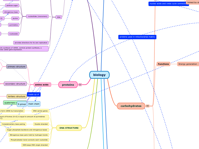
proteins
amino acids
primary structure
main chain
amino group
carbonyl group
peptide bonds
secondary structure
main chain
beta pleated sheets
alpha helices
tertiary structure
interactions of R groups
quaternary structure
DNA STRUCTURE
DNA carries genes
Used to form mRNA by transcription
mRNA used to form proteins through translation
Amount of Purines (A+G) is equal to amount of pyrimidines (T+C)
Double stranded
Complementary base pairing
Sugar phosphate backbone and nitrogenous bases
Nitrogenous base pairs held by hydrogen bonds
Phosphodiester bond connects each nucleotide
SSB keeps DNA single stranded
Additional info
Additional info
R group
main chain
hydrophobic interactions
ionic bonds
hydrogen bonds
Van der Waals interactions
covalent bonds
polypeptide
DNA expression
Prokaryotes
(transcription and
translation coupled)
RNA polymerase (RNAP)
70 s ribosome
f MET first starting amino acid
Transcription
(Starting point +1)
Nucleotides in DNA to the right are labeled by positive numbers and this area of the DNA is referred to as downstream. To the left of the transcription start site nucleotides are numbered by negative numbers and this is called as the upstream
INITIATION
RNA polymerases bind to a region on the DNA upstream of the start site. This region is called the promoter. one direction only 5’ to 3’. The enzyme binds the promoter and picks one of the two strands as the template to use to form mRNA. It reads the template in 3’ to 5’ direction and forms mRNA in 5’ to 3’ direction.
ELONGATION
new nucleotides being added to the 3’ end of the new strand. The original strand is called as the template strand.
TRANSLATION
First, a correct match between a t R N A and an amino acid, done by the enzyme aminoacyl-t R N A synthetase, Second, a correct match between the t R N A anticodon and an mRNA codon. Flexible pairing at the third base of a codon is called wobble and allows some tRNA’s to bind to more than one codon
. The RNA is called ribosomal RNA (or rRNA). Each ribosome is made of two parts or subunits – large and small. These are typically separated, but come together when translating an mRNA.
E SITE
the exit site, where discharged tRNA’s leave the ribosome
P SITE
holds the tRNA that carries the growing polypeptide chain
translation brings together mRNA, tRNA bearing the first amino acid of the polypeptide, and two subunits of a ribosome
The mRNA is read in 5’ to 3’ direction
while the amino acids are added from
N to C direction
A SITE
site holds the tRNA that carries the next amino acid to be added to the chain
TERMINATION
Once a stop codon is reached in the A site there is no tRNA that corresponds to a stop codon. Instead a Release factor sits in the A site, dissociating the complex stopping translation. This is a GTP driven process.
Eukaryotes
(transcription and
translation NOT coupled)
RNA polymerase II – pre mRNA,
snRNA, microRNA
dn
signal for the cell to make a cut in the
newly formed pre mRNA and release
it from DNA. At the 5’ end a modified
G nucleotide is added, this is called
CAP and at the 3’ end near the AAUAAA
sequence a polyA tail is added by the
enzyme polyA polymerase. The 5’cap
will be used for translation and the
3’polyA tail helps with stability of the mRNA.
RNA SPLICING
Before the pre mRNA exits the nucleus
to be translated it has to go through
RNA processing. This process involves
removal of introns and joining togethe
r of exons. ONLY IN EUKARYOTES
ALTERNATE SPLICING
to form different mRNAs and hence different proteins.
80 S ribosome
MET first starting amino acid
Floating topic
DNA STRUCTURE
DNA carries genes
Used to form mRNA by transcription
mRNA used to form proteins through translation
Amount of Purines (A+G) is equal to amount of pyrimidines (T+C)
Double stranded
Complementary base pairing
Sugar phosphate backbone and nitrogenous bases
Nitrogenous base pairs held by hydrogen bonds
Phosphodiester bond connects each nucleotide
SSB keeps DNA single stranded
DNA REGULATION
nucleosome
linker DNA
histones
H1
H2A
H2B
H3
H4
Eukaryotes
transcription factors
general
background/basal
expression
specific
activators
INCREASE levels of
transcription
repressors
REDUCE levels of
transcription
control elements
proximal
distal
Prokaryotes
operons
operators
positive regulation
activator present
transcription occurs
activator NOT present
transcription does
NOT occur
negative regulation
repressor present
transcription does
NOT occur
repressor NOT present
transcription occurs
differential gene expression
CELL SIGNALING
signal molecule
ligand
signal
long distance signaling
receptor
intracellular receptor
membrane receptor
G Protein Coupled Receptor
signal molecule
GPCR
G protein
GDP
GTP
activated G protein
enzyme
cellular response
GTP
G protein
GDP
Tyrosine Kinase Receptor
signal molecule
receptor tyrosine
kinase receptor
dimer
activated tyrosine
kinase regions
ATP
ADP
fully activated
tyrosine kinase
receptor
inactive relay proteins
phosphate groups
activated relay proteins
cellular response
Ion Channel Receptor
ligand-gated ion
channel receptor
signal molecule
channel opens
specific ions to
flow through
cellular response
signal molecule
channel closes
local signaling
paracrine signaling
signal binds to receptors
stimulate adjacent cells
synaptic signaling
occurs between cells
with synapses
cell releasing signal is close to target cell
stages
reception
signal molecule binds to receptor
transduction
relay molecules
response