por Michelle Bordalba hace 23 días
35
Unit 4 - Animal Systems

por Michelle Bordalba hace 23 días
35

- proteins that speed up biochemical reactions such as hydrolysis - Hydrolysis: chemical reactioin where water breaks macromolecules into smaller molecules
structural support for cellulose & chitin
long term energy
many monosaccharides
maltose, sucrose (table sugar)
short-term energy
2 monosaccharides
3 to 7 carbons in a C : H : O ratio of 1 : 2 : 1
glucose, fructose
instant energy
recoil of blood against closed semilunar valves
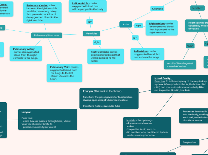
recoil of blood against closed AV valves
Right atrium: carries deoxygenated blood that is pumped to the right ventricle
Left atrium: carries oxygenated blood that comes from the lungs
Ventricles
Right ventricle: carries deoxygenated blood that will be pumped to the lungs
Left ventricle: carries oxygenated blood that will be pumped to the body
Pulmonary Structures
Pulmonary Valve: valve between the right ventricle and the pulmonary artery that prevents backflow of deoxygenated blood to the right ventricle.
Pulmonary Vein: carries oxygenated blood from the lungs to the left atriums towards the heart.
Pulmonary Artery: carries deoxygenated blood from the right ventricle to the lungs.
Vena Cava
Inferior Vena Cava: carries deoxygenated blood from the lower body to the right atrium
Superior Vena Cava: carries deoxygenated blood from the upper body to the right atrium
Aorta & Aortic Valves
Aortic valve: prevents backflow of oxygenated blood to the left ventricle. Located between the left ventricle and the aorta.
Aorta: carries oxygenated blood from the left ventricle to the rest of the body
Capillaries
- not elastic - no valves - permeable - large lumen - very low pressure - flows slowly - most abundant
site of gas exchange with tissue cells
Veins
- little elasticity - valves - low permeability - large lumen - low pressure -flows slowly - largest: vena cava, smallest venules
carries blood towards the heart
The Stomach: Structure - the stomach has 3 regions (cardiac, fundic, pyloric) & 2 sphincters at each end (cardiac & pyloric sphincters.) Function: food is mixed with gastric juices and churned, preparing the chyme (semifluid mass when food is mixed with digestive juices) for further digestion in the small intestine.
Small Intestine: Structure: Long, tiny tubes that take up most of the lower abdomen. made up of 3 main sections. Function: break down food and absorb nutrients Duodenum: where digestive juices and enzymes are produced and used to break down food. Jejunum: It's where food is mixed with stomach acid. Ileum: Nutrients are absorbed from the digested food then waste is moved to the large intestines.
Large Intestine: Structure: made up of 3 sections Function: Ascending Colon: Absorbs water and electrolytes from undigested food. Transcending Colon: Moves food waste toward the descending colon. Descending Colon: Continues the process of turning food into feces.
Mechanical Digestion: breaking food into smaller pieces and the physical movement of food
Larynx: Function: - voice box, air passes through here, where your vocal cords vibrate to - produce sounds (your voice)
Trachea: Function: It is the tube that carries air from your mouth and nose down to your lungs, helping you breathe Structure: - Made of C-shaped cartilage rings that keep the trachea open and allow airflow. - Lined with cilia, tiny hair-like structures that help push mucus and debris out of the lungs.
Bronchus (pl. Bronchi) - The 2 air tubes that connect the trachea to the lungs - The trachea “forks” into the 2 bronchi
Bronchioles: Function: Small branches that come from the bronchi and lead to the tiny air sacs in the lungs called alveoli Structure: surrounded by smooth muscles that contract or relax to control the size of the airways.
Alveoli: Function: The alveoli are where gas exchange occurs in the lungs. It provides a large surface area for gas exchange. Structure: Tiny, balloon-like sacs at the end of the bronchioles. Clustered together like bunches of grapes.
The Lungs Function: Responsible for breathing. Takes in oxygen from the air, remove carbon dioxide from the blood and helps oxygenate the body. Structure: A nonmuscular organ filled with bronchial tubes, bronchioles, and alveoli for gas exchange. It’s also surrounded by pleural membranes and protected by the rib cage.
Diaphragm Function: A dome-shaped muscle under the lungs that helps with breathing by moving down to let the lungs expand and up to push air out. Structure: Large, flat muscle that separates the chest from the abdomen. It’s located beneath the lungs.
Carbon Dioxide Transport at the Tissue Level
In the tissues, carbon dioxide (CO2) moves from the tissues into the red blood cells (attached to hemoglobin).
The deoxygenated blood travels to the heart.
The heart pumps the deoxygenated blood (now with CO2) to the lungs.
In the lungs, CO2 diffuses from the red blood cells into the alveoli.
CO2 is exhaled out of the body.
Oxygen Transport at the Tissue Level
Oxygen from the air diffuses from the alveoli into red blood cells in the capillaries.
The oxygenated blood travels from the lungs to the heart.
The heart pumps oxygen-rich blood to the body's tissues.
Oxygen diffuses from the red blood cells into the tissues, providing them with oxygen.
Expiration
- The diaphragm moves upward & relaxes - The rib cage moves in and out - The volume of chest cavity decreases - Air pressure increase
Inspiration
- Diaphragm moves downward & contracts - Rib cage moves up and out - Volume of chest cavity increases - Air pressure lowers