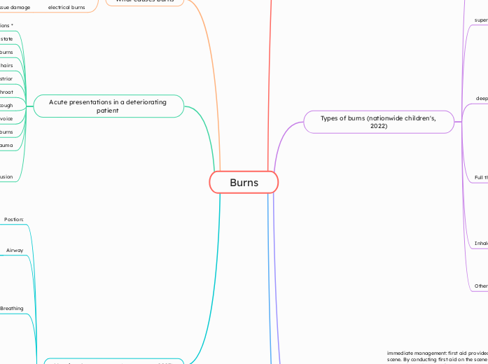
Burns
Nursing Asessments: (treatment, 2023)
Exposure
skin inspection
apply cling wrap to burn
remove all non-adhered clothes and jewelry
temperature: if less than 35.5 provide blacnkets and warm the patinet
Disability:
Assess pain
if indicated, provide analgesia
Conducte a GCS score, pupillary response and limb strength
if reduced level of consciouness, consider shock or hypoxaemia
Circulation:
Look out for signs of shock
hypotension
abnormal skin colour
tachycardia
check blood pressure and pulse and cardiac rhythm
colour and warth of skin, ensure patient stays warm due to risk of hypoxia
perfusion and capillary refill
Breathing
severe burns: apply oxygen through non-rebreather mask
maintain O2 sats
Auscultate chest: listen for chest sounds like stridor to see if their is anything inhaled
respiratory rate: assist ventilation if required
Airway
evidence of airway burn: give humidified oxygen via non-rebreather. Be quick with oxygen as airway injuries can worsen overtime. Consider and prepare for intubation
Patency: maintain the airway patency
Postion:
comfot for the patient. patients with head and neck burns should be head up to reduce swelling
Acute presentations in a deteriorating patient
Changes in perfusion
skin: warm, red and tender with little to no sensation
pulse pressure:
capillary refill increased
burns with trauma
mid-deep dermal or full thickness burns
hoarse voice
cough
sore throat
strior
singed faical hairs
inhalation, facial or mouth burns
reduced conscious state
clincial manifestions on each presentations *
What causes burns
electrical burns
deep tissue damage
potnetial for internal organ damage
lead to cardiac arrest or arrythmias
chemical burns
cause more severe tissue damage
pain, redness, tissue necrosis
Thermal burns
causes redness, blistering and swelling
ranges from superficial to fullthickness burns
inhalation injuries
Nursing Inteverventions (Zwierello et al, 2023)
Overall nursing interventions:
Assess pain:
administer pain relief
after reciving a burn, patient is going to be in alot of pain, if patient is consious ask patient to rate thier pain out of 10 and document it
wound:
apply cling wrap on the new burn after cleaning it
remove necotric tissues to prevent any infections
Becuase the extent of the burns can cause a severity of fluid loss
fluid: administer IV fluids
Prority assessments:
fluid balance: assess urine output
Circulation: monitor bp, hr and capillary refill
Airway: check for soot in air way, strider present
Management
State wide interventions: (Alfred Health, 2025)
The Alfred Health Victoria Adult Burns service provides statewide burns care, offers clinical practice guidelines, long term follow up and rehabilitation
provides resources and guidelines for burns assessment and management
minor burn care can be completed without being in hospital and clinics with the appropriate wound management
Burn prevention: education and awareness
Pharmacological interventions (uptodate, 2025)
hyperglyceamia can occur after a burn
check BGL
may require medications like metformin
might require high flow oxygen
Fluid resuscitation: required IV to prevent dehydration and organ failure
Antibitotics and antimicrbiols to prvent and treat wound infections
Analgestics: combination of pain relief
immediate management: first aid provided at the scene. By conducting first aid on the scene can prevent burns from getting severly worse, howver depends on the extent of the injury to what first aid is completed. (McCann et al, 2022)
Chemical burns: patient to be removed from contaminated clothing, irrigated with running water to remove chemical from burn. Irrigated fro 45 mins- 1 hr (McCann et al, 2022)
Warm the patient: by warming the patinet is can prevent the possibility of hypothermia (McCann et al, 2022)
Cover the burn with non-adhernt dressing such as cling wrap. (McCann et al, 2022)
Running under cool water: to be completed for 20 mins even up to 4 hours after injury. this way the tissue damage is arrested and wounds wouldnt be as deep as it could of been (McCann et al, 2022)
Types of burns (nationwide children's, 2022)
Other
infection
shock
dehydration
Inhalation injuries
stridor
coughing
difficualty breathing
Full thickness burns:
blisters: epidermis & dermis destroyed no blisters
colour: white/charred/black
sensation: no sensation
appearance: white waxy charred, no blisters, no cappliary refill
pathology: involves epidermis, dermis destroyed
deep dermal burns:
colour: white/pale pink/ blotchy red
circulation: sluggish capillary refill
Sensations: decreased sensations
Appearance: blotchy red or pale deeper dermis
pathology: involves epidermis and significant part of dermis
superficial dermal thickness burns:
colour: pink
circulation: hypomanic, rapid capillary refill
Sensation: tender and painful, increased sensation
Appearance: pale pink, smaller blisters, wound base blaches with pressure
Pathology: involves dermis and epidermis
Superficial epidermal burns:
colour: red and warm
circulation: normal
Sensation: might be painful
Appearnace: dry and red, no blisters
Pathology: expidermis only
What is a burn
burns falls under the shock category. Shock is a complex life threatening condition that comes from circulatory failure. As a result of severe burns, the body falls into a state where it is unable to deliver sufficient oxygen to the cells and tissues (Blumlein & Griffins, 2022)
multi-organ failure: (Jeschke et al, 2020)
immune suppression (uptodate, 2025)
leads to patients being more susceptible to causing infections
increases risk of sepsis
imbalances in fluid
loss of fluid increases capillary permability
can lead to hypovalemia
overall damages kidney and heart
severe burns can lead to infection (feng et,al, 2028)
increase risk of sepsis
bodys reaction to infection is damges its own tissues and organs
tissue hypoperfusion:
burn injuries lead to increased capillary permermibility
lead to decrease in BP, and impaired tissue perfusion
severe burns causes a significant loss of fluid
fluid loss leads to decrease in intravascular volume
Results in hypoxia: as a result of poor oxygen supply via circulation
burns can cause a build up of fluid in the burn area which can restrict oxygen delivery
large burns can lead to hypovolemia, impairing oxygen delivery to tissues
severe burns can damage blood vessels
burns are a injury to the skin or tissue caused by
heat, electricity or other sources. Burns vary in
severity.
Systematic Response:
Metabolic changes: metabolic rate increases up to three times its orignial rate (the royal childrens hospital melbourne, 2024)
Respiratory changes: cause bronchoconstricton occurs (the royal childrens hospital melbourne, 2024)
Cardiovascular changes:
capillary permeability increased, loss of fluids in intestinal compartment. Myocardial contractibility is decreased. Results in hypotension (the royal children's hospital Melbourne, 2024)
Local response:
Zone of hyperaemia: outermost zone tissue perfusion increased (the royal childrens hospital melbourne, 2024)
Zone of stasis: decreased tissue perfusion,
tissue is potentially salvageable (the royal childrens hospital melbourne, 2024)
Additional impacts:
- prolonged hypotension
- infection risk
- oedema
Zone of coagulation: irreversible tissue loss
due to coagulation (the royal childrens hospital melbourne, 2024)

