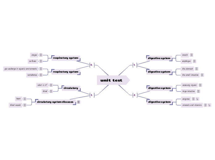
unit test
circulatory system diseases
Coronary heart disease
- Coronary arteries provide blood to the heart tissue itself
- Sometimes, these can become partially blocked with plaque (fat, cholesterol, calcium and other substances usually found in the blood)
- Can be caused by genetic factors, as well as lifestyle factors (unhealthy diet, smoking, lack of exercise)
- Symptoms include tiredness, diziness, and pain in the chest and arms
- It can be diagnosed using an angiogram: dye is injected into the artery and an x-ray is taken
Heart attack:
- If a coronary artery is completely blocked (plaque or a blood clot), the heart muscle will not receive oxygen; the heart will stop functioning - this is a heart attack
- Symptoms include pain, difficulty breathing, nausea, sweating, dizziness
- Heart attacks can be diagnosed with an ECG (electrocardiogram)- this measures the electrical activity of the heart and can help identify is there is any damaged heart tissues
blood vessels
- There are three types of blood vessels: arteries, veins, and capillaries
- Arteries: thick-walled blood vessel that carries blood away from the heart
- The walls of arteries are thick to withstand the pressure of the heart
- Veins: a blood vessel that returns blood to the heart
- Blood in veins is at a lower pressure so the walls of veins do not have to be as thick and elastic as arteries
- Capillaries: tiny thin-walled blood vessel that enables the exchange of gases, nutrients, and wastes between the blood and the body tissues
- Oxygen, nutrients and waste diffuse (move between) the cells of the body and blood through these thin capillaries
heart
- Heart is made of three types of tissues: cardiac muscle tissue, nerve tissue
- The contraction of the cardiac muscle causes the blood to move around the body
- There are valves in the heart that control the direction of blood flow- this is what you hear when you hear a heartbeat
- Your heart rate (pulse) changes depending on things like stress, physical activity, and temperature
- The heart muscle is covered by smooth epithelial tissue
circulatory
blood
- Blood is a connective tissue
- Blood flows through the lungs to pick up oxygen and then flows through the body and delivers it to active cells.
- Blood also carries waste away from the cells
- Carbon Dioxide: Blood also carries carbon dioxide to the lungs where it is exhaled
Other waste: Blood also carries other waste to the kidneys; the kidney filters these wastes out of the blood and excretes them in the form of urine
- There are four types of blood cells:
- Red blood cells: most plentiful in the blood, they have a protein called hemoglobin (which gives red colour), this protein carries oxygen throughout the body
- White blood cells fight infection; they make up only 1% of blood cells
- Platelets: tiny cells that help with blood clotting; less than 1% of blood cells
- Plasma: is protein-rich liquid that carries the blood cells along; makes up half of blood volume
what is it?
Circulatory system: the organ system that is made up of the heart, the blood, and the blood vessels; this system transports oxygen and nutrients throughout the body and carries away wastes
- The circulatory system also helps regulate body temperature and fight pathogens (like bacteria, viruses, etc.- this is called the immune system)
3 parts
- Blood
- The heart
- Blood vessels
vertebrates
- Air does not just flow into and out of the lungs on its own.
- The brain acts as a respiratory control centre to coordinate breathing movements and regulate the breathing rate. It also monitors the volume of air in the lungs and the gas levels in the blood.
- Two sets of structures—the muscular diaphragm and the rib muscles—control the air pressure inside the lungs.
- Changes in air pressure cause air to move into and out of the lungs.
- The diaphragm is a dome-shaped layer of muscle that separates the region of the lungs (the thoracic cavity) from the region of the stomach and liver (the abdominal cavity). The rib muscles, or intercostal muscles, are found between the ribs and along the inside surface of the rib cage.
gas exchange in aquatic environments
- Aquatic environments contain oxygen in the form of dissolved gas. Many aquatic organisms, take in oxygen through gills.
- Gills are physical adaptations that enable organisms to carry out gas exchange in aquatic environments.
- A fish exchanges gases by taking water into its mouth and ventilating (or pumping) it over the gills. As water flows across the gills, dissolved oxygen in the water goes into the fish’s blood. At the same time, carbon dioxide goes out of the blood, across the gill tissue, into the water and is carried out of the fish’s body when the water passes out of the gill openings.
- Another adaptation used by fish is a countercurrent exchange mechanism.
- Blood and water flow through the gills in the opposite directions. Oxygen diffuses along a gradient, called a diffusion gradient, meaning that the oxygen molecules move from a region of high concentration to a region of low concentration. This countercurrent exchange mechanism keeps the diffusion gradient of the oxygen high.
respiratory system
surfaces
there are 2 main requirements for respiration
- 1. Respiratory surface (the area of an animal’s body where gases are exchanged with the environment) must be large enough for the exchange of oxygen and carbon dioxide to occur quickly enough to meet the body’s needs.
- 2. Respiration must occur in a moist environment so that the oxygen and carbon dioxide are dissolved in water
- Ventilation is the process of moving an oxygen-containing medium (water or air) over the respiratory surface (such as the gills, trachea, or lungs).
stages
- The first stage in respiration, breathing, involves two basic processes: inspiration (breathing in, or inhaling) and expiration (breathing out, or exhaling).
- The second stage of respiration, external respiration, is the exchange of oxygen and carbon dioxide between the inspired air inside the lungs and the blood. This stage of the respiratory process performs the vital function of gas exchange. Gas exchange is the delivery of oxygen from the lungs to the blood, and the elimination of carbon dioxide from the blood to the lungs
- The third stage, called internal respiration, is the exchange of oxygen and carbon dioxide between the blood and the body’s tissue cells
- The fourth and final stage in human respiration is cellular respiration. As you have learned, cellular respiration is the series of energy-releasing chemical reactions that take place within the cells. It is the sole means of providing energy for all cellular activities.
minerals and vitamins
enzymes
- Enzymes are important in the process of transferring signals from one cell to another cell. In this process, a chemical or physical signal is transmitted through a cell which is governed by a series of molecular events and then a cellular response takes place.
- In the process of digestion, enzymes help break down large molecules into smaller molecules because many nutritional contents like sugars, fat, and protein are present in large molecules and cannot be taken up by the human body therefore they are needed to be broken down.
- Enzymes are widely used in the field of biotechnology as molecular scissors to cut DNA fragments or as polymerases to add DNA/RNA fragments.
- Enzymes are also responsible for the generation of movement in the human body with the help of myosin which hydrolyzes ATP to generate energy.
large intestine
- large intestine is also called colon
- large intestine where water is absorbed back into bloodstream and cells
- bacteria break down undigested food further this is where the smell comes from
- some produce important vitamins
- left over material is fees that is excreted
accessory organs
- accessory agar's are connected to the digestive system by ducts fluids produced by accessory organs are essentials for digestion
- pancreas pancreatic fluid contains enzymes that chemically digest carbohydrates, lipids and proteins
- also they contain bicarbonate a base that an change the chyme xx
the small intestine
- the small intestine digestion is completed here macromolecules are broken down into their component molecules
- nutrients are absorbed through the membranes that line that small intestine where they pass the circulatory system
- the circulatory system carries the nutrients to the rest of the body
- there are 3 parts of the small intestine duodenum, jejunum and ileum
the duodenum
- short, wide u shaped section of the small intestine where food travels after leaving the stomach
- the walls of duodenum recovered in villi
- each villi are covered in microvilli which is also increased the surface area
jejunum and ileum
- ileum the portion of the small intestine that follows immediately after the jejunum
- contains more folds that duodenum
- breaks down remaining proteins and carbohydrates
- the portion of the small intestine that follows immediately after jejunum
- contains fewer and smaller villi to absorb nutrients
- pushes material into large intestine
the stomach
- the stomach muscular j shaped organ where mechanical digestion happens
- contains gastric juice a mixture of hydrochloric acid, salts,enzymes,water,mucous that is produced by the stomach to help digest food
- mucous coats the walls of the stomach to protect it from the gastric juices
- pyloric sphincter controls the flow of chyme into small intestine
digestive system
esophagus
- esophagus the muscular tube throughout which food passes from the mouth to the stomach
- epiglottis prevents food from going down the wrong tube so no food gets in ur lungs
- peristalsis a wave like a series of muscular contractions in the esophagus
mouth
- salivary glands produce saliva to begin the chemical digestion
- saliva contains amylase (enzyme) that breaks carbohydrates
- saliva helps us taste the flavours of food
- mechanical digestion also happens in the mouth teeth bite and tear area
- this also increases the surface area for chemical digestion